Paranasal Sinuses
Acute Bacterial Sinusitis
A bacterial infection of the paranasal sinuses that most often develops as a complication of a viral upper respiratory infection. Diagnosis is clinical and depends primarily on the pattern and trajectory of symptoms.
Clinical Vignette
The cold was getting better—so why is she sick again?
Recognize worsening after initial improvement as one of the characteristic clinical presentations of acute bacterial sinusitis.
An 8-year-old presents with worsening nasal congestion, nasal discharge, and cough. Eight days ago, she developed rhinorrhea, sore throat, and a mild cough consistent with a viral upper respiratory infection.
By illness day 5, her symptoms seemed to be improving and she had returned to normal activity. Over the last 2 days, however, her nasal congestion and daytime cough have become substantially worse, and she developed a new fever to 39.1°C (102.4°F).
Examination shows mucopurulent nasal discharge and edematous nasal mucosa. She is breathing comfortably with normal oxygen saturation and clear lungs.
Her parent asks whether this is simply the same cold taking longer than expected.
Your clinical reasoning task
Does this still fit the expected trajectory of an uncomplicated viral URI, or does the initial improvement followed by new fever and worsening respiratory symptoms suggest acute bacterial sinusitis?
Anatomy & Localization
Localize sinusitis to the paranasal sinuses
Sinus anatomy changes throughout childhood, and the location of infection helps predict both symptoms and complications.
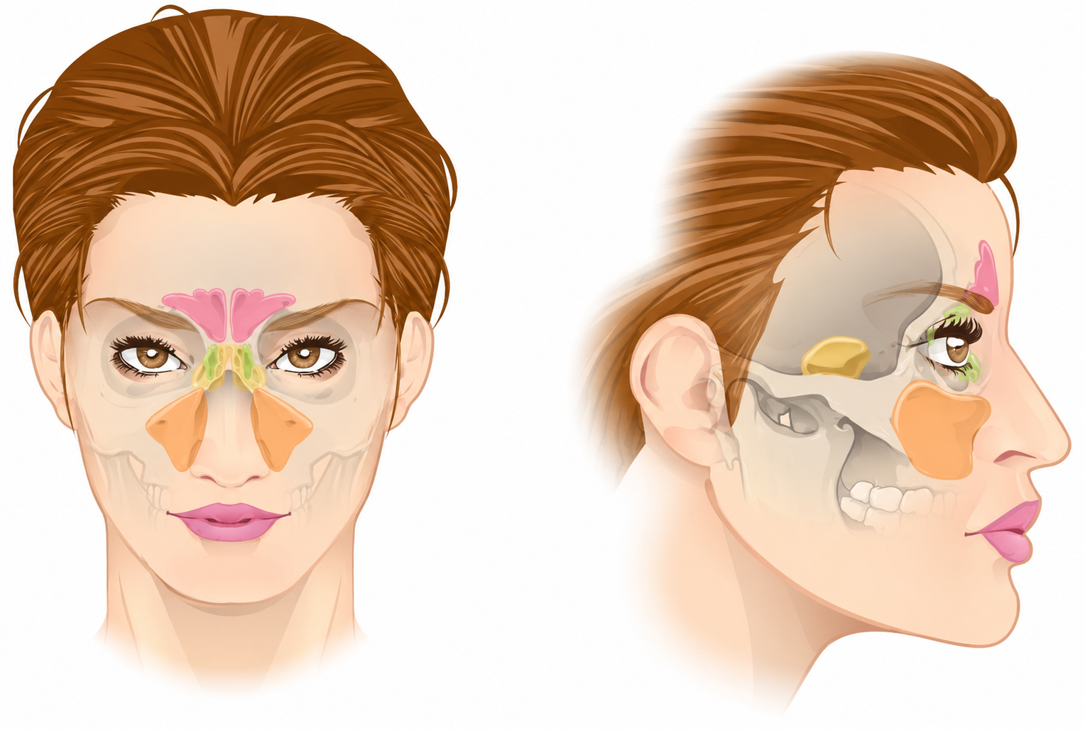
Paranasal sinus anatomy shown in anterior and lateral views. The frontal, ethmoid, sphenoid, and maxillary sinuses are highlighted to demonstrate their relative location and relationships to surrounding structures.
Adapted from OpenStax College, Anatomy & Physiology, licensed under CC BY 3.0. Modified for PediAtlas.
Maxillary & Ethmoid Sinuses
The maxillary and ethmoid sinuses begin developing during fetal life and are present, although small, at birth. They continue to enlarge throughout childhood.
Frontal & Sphenoid Sinuses
The frontal and sphenoid sinuses develop later, typically beginning around 2–3 years of age, and remain relatively rudimentary during early childhood.
Development
Sinus anatomy changes with age
Middle Meatus
The maxillary, frontal, and anterior ethmoid sinuses drain into the middle meatus. Inflammation and edema around these drainage pathways can obstruct sinus outflow and impair clearance of secretions.
Superior Meatus
The sphenoid and posterior ethmoid sinuses drain more posteriorly through the superior meatus, providing a separate drainage pathway into the nasal cavity.
Clinical Anatomy
Why drainage matters
Complication Risk
Anatomy predicts complications
Pathophysiology
A viral URI sets the stage for bacterial sinusitis
Acute bacterial sinusitis usually develops when inflammation from a viral upper respiratory infection disrupts normal sinus drainage and mucociliary clearance.
Viral URI
A respiratory virus infects the nasal and sinus mucosa, triggering inflammation and increased mucus production.
Mucosal edema
Inflammation causes swelling of the respiratory mucosa surrounding the narrow sinus drainage pathways.
Ostial obstruction
Edema narrows or obstructs the sinus ostia, reducing normal ventilation and drainage of the affected sinus.
Impaired clearance
Obstruction and impaired mucociliary function allow secretions to accumulate within the sinus rather than being cleared normally.
Bacterial infection
The obstructed, poorly draining sinus creates conditions that allow bacteria to proliferate, producing acute bacterial sinusitis.
Core Mechanism
The problem begins with impaired drainage
Common Pathogens
Think pneumococcus, H. influenzae, and Moraxella
Clinical Translation
This is why the trajectory matters
History & Physical
The diagnosis is in the timeline
Acute bacterial sinusitis is primarily a clinical diagnosis. Focus on how symptoms began, how long they have lasted, and whether the child is improving or worsening.
Diagnostic Framework
Recognize one of three characteristic patterns
1. Persistent Illness
>10 days without improvement
Nasal discharge of any quality, daytime cough, or both persist for more than 10 days without appreciable improvement.
The key is not simply reaching day 10—it is reaching day 10 without a meaningful trend toward improvement.
2. Severe Onset
≥39°C + purulent discharge
High fever of at least 39°C (102.2°F) occurs together with purulent nasal discharge for at least 3 consecutive days at the beginning of the illness.
Fever and purulent discharge must occur together—the combination and duration distinguish this pattern from an uncomplicated viral URI.
3. Worsening Course
Improve → then worsen
A child with an apparent viral URI begins to improve and then develops substantial worsening of nasal discharge, nasal congestion, or daytime cough, or develops a new fever.
This biphasic pattern is often called “double sickening.”
Common Trap
Green mucus does not equal bacterial sinusitis
Symptoms to Ask About
- • Duration and trajectory of nasal symptoms
- • Nasal congestion or obstruction
- • Nasal discharge of any quality
- • Daytime cough, which may worsen at night
- • Fever and when it occurred during the illness
- • Initial improvement followed by worsening
- • Headache or facial discomfort
- • Decreased appetite, fatigue, or halitosis
Physical Examination
- • General appearance and vital signs
- • Nasal discharge and congestion
- • Nasal mucosal or turbinate swelling
- • Periorbital swelling or erythema
- • Extraocular movements and ocular pain
- • Visual changes or proptosis
- • Facial or forehead swelling
- • Neurologic status when complications are suspected
Physical Exam
There is no single exam finding that confirms uncomplicated ABS
Red Flags
Look beyond uncomplicated sinusitis
Differential Diagnosis
Not every prolonged runny nose is sinusitis
Many common pediatric conditions produce nasal discharge and cough. Distinguish them by the symptom trajectory, associated findings, and whether the illness fits one of the characteristic patterns of acute bacterial sinusitis.
Viral Upper Respiratory Infection
The most important mimic. Viral URIs commonly cause nasal congestion, cough, fever, and even thick or purulent nasal discharge.
Symptoms generally begin improving within about 10 days. Acute bacterial sinusitis becomes more likely when symptoms persist without improvement, are severe from the onset, or worsen after initial improvement.
Allergic Rhinitis
Consider allergic rhinitis when symptoms are recurrent or chronic and include sneezing, nasal itching, clear rhinorrhea, or other features of atopy.
The characteristic persistent, severe, or worsening illness pattern of acute bacterial sinusitis is usually absent.
Nasal Foreign Body
Particularly important in younger children. A retained nasal foreign body classically produces persistent, unilateral, foul-smelling or purulent nasal discharge.
Unilateral symptoms should prompt careful examination of the nasal cavity rather than automatically attributing the discharge to sinusitis.
Adenoiditis / Adenoid Hypertrophy
Adenoid disease can produce nasal obstruction, persistent nasal discharge, mouth breathing, snoring, and cough, substantially overlapping with symptoms of sinusitis.
Chronic or recurrent symptoms without a characteristic acute sinusitis trajectory should raise consideration of adenoid disease.
Pertussis
Consider pertussis when cough becomes the dominant feature, particularly with prolonged paroxysms, inspiratory whoop, post-tussive emesis, or known exposure.
The respiratory history may begin with nonspecific upper respiratory symptoms before the characteristic cough develops.
Lower Respiratory Infection
Pneumonia or bronchiolitis can also produce cough and fever. Tachypnea, hypoxemia, increased work of breathing, focal lung findings, or prominent lower respiratory symptoms should shift attention below the upper airway.
Clinical Shortcut
Start by asking one question
Diagnostic Workup
Uncomplicated sinusitis is a clinical diagnosis
Most children with acute bacterial sinusitis do not need laboratory testing, cultures, or imaging. Diagnostic testing becomes important when complications or an alternative diagnosis are suspected.
Uncomplicated ABS
The history usually makes the diagnosis
Laboratory Testing
Not routinely indicated.
CBC, inflammatory markers, and other routine laboratory studies are generally unnecessary for uncomplicated acute bacterial sinusitis.
Cultures
Do not routinely obtain nasal cultures.
Cultures of nasal secretions do not reliably identify the organisms causing infection within the paranasal sinuses and are not recommended for routine diagnosis.
Imaging
Do not image uncomplicated ABS.
Plain radiographs, ultrasound, CT, and MRI should not be used simply to distinguish acute bacterial sinusitis from an uncomplicated viral URI.
Why Not Image?
Abnormal sinuses do not necessarily mean bacterial sinusitis
Suspected Extension
Orbital or intracranial findings change the workup
Obtain urgent advanced imaging when the examination raises concern for orbital or intracranial complications.
Concern for orbital extension
- • Proptosis
- • Painful or restricted extraocular movements
- • Visual changes
- • Significant periorbital swelling or chemosis
Concern for intracranial extension
- • Severe or progressive headache
- • Vomiting
- • Altered mental status
- • Focal neurologic findings
- • Seizure
- • Forehead swelling or tenderness
Advanced Imaging
Use contrast-enhanced CT and/or MRI when complications are suspected
Management
Treat based on the clinical pattern
Management depends on whether the child has persistent illness, severe onset, or a worsening course—and on the risk for resistant organisms or complications.
Persistent Illness
Antibiotics OR brief observation
For symptoms lasting more than 10 days without improvement, either begin antibiotic therapy or observe for up to 3 additional days if the child is otherwise well and reliable follow-up is available.
Start antibiotics if the child worsens at any time or fails to improve during the observation period.
Severe Onset
Start antibiotics
Children with fever ≥39°C (102.2°F) and purulent nasal discharge for at least 3 consecutive days at the beginning of illness should receive antibiotic therapy rather than observation.
Worsening Course
Start antibiotics
Children who initially improve from an apparent viral URI and then substantially worsen or develop a new fever should receive antibiotic therapy.
First-Line Therapy
Amoxicillin with or without clavulanate
Standard-Dose Amoxicillin-Clavulanate
45 mg/kg/day
Dose according to the amoxicillin component, divided twice daily.
Maximum amoxicillin component: 1.75 g/day.
Appropriate for many children with uncomplicated ABS who do not have risk factors for resistant organisms.
High-Dose Amoxicillin-Clavulanate
90 mg/kg/day
Dose according to the amoxicillin component, divided twice daily.
Maximum amoxicillin component: 4 g/day.
Use for severe disease or when there is increased risk for resistant organisms.
Resistance Risk
When should you consider high-dose therapy?
When is amoxicillin alone reasonable?
Amoxicillin alone may be considered for mild, uncomplicated disease when the child has no risk factors for resistant organisms and local prevalence of β-lactamase–producing respiratory pathogens is low.
Amoxicillin-clavulanate provides additional activity against β-lactamase–producing organisms such as H. influenzae and M. catarrhalis.
Reassessment
Expect improvement within 48–72 hours
Supportive Care
- • Maintain adequate hydration
- • Acetaminophen or ibuprofen for pain and fever when appropriate
- • Saline nasal irrigation may provide symptomatic relief
Not Routinely Recommended
- • Routine antihistamines for uncomplicated ABS
- • Routine oral or topical decongestants
- • Routine mucolytics
Evidence supporting these adjunctive therapies in uncomplicated pediatric ABS is limited.
Complications
When infection escapes the sinuses
Most children recover without complication, but extension beyond the paranasal sinuses can threaten vision, neurologic function, and life.
Anatomic Spread
Think orbit and brain
Orbital Complications
Orbital involvement is the most common serious complication of acute bacterial sinusitis and commonly results from extension of ethmoid sinus infection.
- • Preseptal / periorbital inflammation
- • Orbital cellulitis
- • Subperiosteal abscess
- • Orbital abscess
Worry about postseptal disease when there is:
- • Proptosis
- • Pain with extraocular movements
- • Restricted extraocular movements
- • Chemosis
- • Decreased visual acuity
Intracranial Complications
Intracranial extension is less common than orbital disease but carries greater potential morbidity. It is particularly associated with frontal sinusitis.
- • Epidural empyema
- • Subdural empyema
- • Brain abscess
- • Meningitis
- • Intracranial venous thrombosis
Red flags include:
- • Severe or progressive headache
- • Vomiting
- • Altered mental status
- • Seizure
- • Focal neurologic findings
Pott Puffy Tumor
Frontal bone osteomyelitis + subperiosteal abscess
Despite the name, this is not a tumor. Extension of frontal sinus infection can produce osteomyelitis of the frontal bone with a subperiosteal abscess.
Suspect it in a child or adolescent with forehead swelling and tenderness in the setting of sinusitis.
Cavernous Sinus Thrombosis
Infection spreads into the cavernous sinus
Infection can reach the cavernous sinus through venous pathways or direct spread, including from sphenoid sinusitis.
Cranial nerve dysfunction can develop; the AAP material highlights lateral gaze paralysis as an early finding.
Clinical Pearl
A swollen eye is not automatically simple periorbital cellulitis
Emergency Escalation
Complicated sinusitis requires hospital-level care
Disposition
Most children can be managed at home
Disposition depends on clinical appearance, ability to tolerate outpatient therapy, reliability of follow-up, and whether there is concern for extension beyond the sinuses.
Outpatient Management
Appropriate for most uncomplicated ABS
- • Well-appearing and clinically stable
- • No orbital or neurologic red flags
- • Able to tolerate oral therapy and maintain hydration
- • Reliable caregiver follow-up
Children treated with antibiotics should have clear return precautions and be reassessed if symptoms worsen or fail to improve within approximately 48–72 hours.
Hospital Evaluation / Admission
Escalate when disease may extend beyond the sinuses
- • Suspected orbital complication
- • Suspected intracranial complication
- • Toxic or significantly ill appearance
- • Inability to tolerate appropriate outpatient therapy
- • Clinical deterioration despite treatment
Complicated disease requires hospital-level evaluation, parenteral antibiotics, advanced imaging, and appropriate specialist consultation.
Observation Option
Observation requires reliable follow-up
Return Precautions
Know the findings that should trigger urgent reevaluation
Reassessment
No improvement by 48–72 hours? Reconsider the plan.
Clinical Pearls
What should you remember on your next shift?
High-yield principles for recognizing and managing acute bacterial sinusitis in children.
The diagnosis is in the timeline
Day 10 is not a magic cutoff
Green mucus does not equal bacterial infection
Do not image uncomplicated sinusitis
The anatomy predicts the complication
Persistent disease is the observation exception
Progressive Quiz
Can you recognize and manage acute bacterial sinusitis?
Work through increasingly clinical scenarios that test diagnosis, workup, treatment, and recognition of complications.
Question 1
A 6-year-old has had nasal congestion and daytime cough for 12 days. The symptoms remain essentially unchanged without meaningful improvement. Which clinical pattern of acute bacterial sinusitis does this represent?
Anki Deck
Acute Bacterial Sinusitis Anki Deck
Reinforce the highest-yield concepts from this module with 18 spaced-repetition cards.
Requires Anki. Deck content is intended for educational review and should be used alongside the full PediAtlas module.
Evidence Base
Clinical References
AAP clinical reference for pediatric acute bacterial sinusitis, including diagnostic criteria, antibiotic management, imaging indications, and recognition of orbital and intracranial complications.
Comprehensive review of pediatric sinus development, pathophysiology, microbiology, clinical presentation, diagnosis, treatment, and complications.
Core pediatric reference for paranasal sinus anatomy and development, pathogenesis, microbiology, clinical diagnosis, treatment, and complications.
Educational Disclaimer
PediAtlas is intended for medical education and should not replace institutional guidelines, local protocols, patient-specific assessment, or clinical judgment.