Retropharyngeal Space & Deep Neck
Retropharyngeal Abscess
Retropharyngeal abscess is a deep neck-space infection characterized by suppuration within the retropharyngeal space, occurring most commonly in young children after infection of retropharyngeal lymph nodes draining the upper aerodigestive tract.
Clinical Vignette
When a sore throat becomes a stiff neck
Recognize the evolution from a common upper respiratory illness to a deep neck-space infection.
A 3-year-old boy is brought to the emergency department after several days of rhinorrhea, sore throat, and fever. His parents initially thought he had a routine viral respiratory illness.
Over the past 24 hours, his fever has increased and he has become increasingly irritable with poor oral intake. His parents notice that he cries when they try to move his neck and has started holding his head tilted slightly to one side.
On examination, he is febrile and uncomfortable appearing. He has limited neck extension and resists turning his head. He swallows reluctantly and occasionally allows saliva to pool in his mouth. The visible oropharynx demonstrates erythema but no obvious unilateral tonsillar enlargement or uvular deviation.
He has no stridor at rest and is currently maintaining his oxygen saturation without supplemental oxygen.
Your clinical reasoning task
Why should fever with painful limitation of neck movement and progressive difficulty swallowing raise concern for infection beyond uncomplicated pharyngitis?
Anatomy & Localization
A deep space hidden behind the pharynx
The retropharyngeal space lies immediately posterior to the pharynx. In young children, lymph nodes within this space drain the upper aerodigestive tract and provide an important pathway for infection.
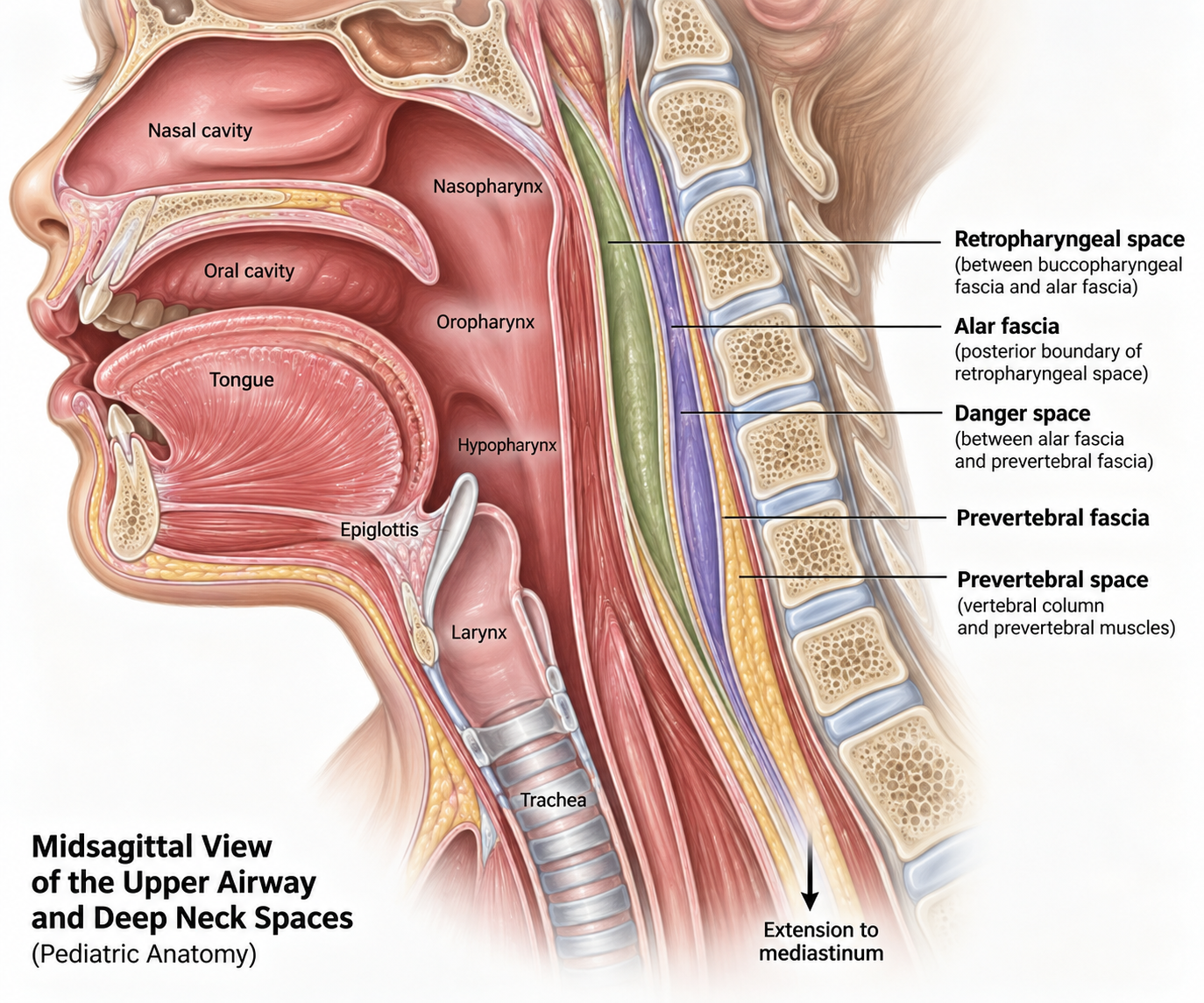
Retropharyngeal space and deep neck anatomy
The retropharyngeal space lies immediately posterior to the pharynx. Infection can extend into adjacent deep neck spaces and descend toward the mediastinum.
Retropharyngeal space
The retropharyngeal space is a potential deep neck space located immediately posterior to the pharynx. It extends from the skull base superiorly into the upper mediastinum inferiorly.
Infection within this confined space can expand anteriorly toward the pharyngeal airway or spread along communicating deep neck spaces.
Retropharyngeal lymph nodes
Young children have prominent retropharyngeal lymph nodes that receive lymphatic drainage from structures including the nasopharynx, adenoids, posterior paranasal sinuses, and middle ear.
Infection from these sites can spread through lymphatic drainage to the retropharyngeal nodes, producing suppurative lymphadenitis that may progress to abscess formation.
Pediatric Anatomy Pearl
Why is retropharyngeal abscess primarily a disease of young children?
Retropharyngeal lymph nodes are most prominent during early childhood and begin to involute around 4–5 years of age. This developmental anatomy helps explain why upper respiratory infections can seed the retropharyngeal space more readily in young children and why spontaneous retropharyngeal abscess is much less common later in childhood.
Anterior: pharynx
The pharyngeal wall lies immediately anterior to the retropharyngeal space. Expansion of infection can therefore produce posterior pharyngeal swelling, dysphagia, drooling, and eventually airway narrowing.
Posterior: deep cervical fascia
Fascial layers separate the retropharyngeal space from deeper neck compartments. Extension beyond these boundaries can allow infection to enter adjacent deep neck spaces.
Inferior: mediastinum
Deep cervical spaces communicate inferiorly with the mediastinum. Descending infection can therefore produce mediastinitis, one of the most serious complications of a deep neck-space infection.
Localization Check
The symptoms make more sense once you know where the infection lives
Fever suggests infection, while painful neck movement and torticollis point toward a deep cervical process. Dysphagia, drooling, or posterior pharyngeal swelling reflect the close relationship between the retropharyngeal space and the pharynx. Respiratory symptoms can emerge as an enlarging infection begins to compromise the airway.
Pathophysiology
A common infection finds a deep pathway
In young children, infection of the upper aerodigestive tract can spread through lymphatic drainage to the retropharyngeal lymph nodes, progressing from inflammation to a mature deep neck-space abscess.
An upper aerodigestive infection begins
Infection of structures draining to the retropharyngeal lymph nodes—such as the nasopharynx, adenoids, paranasal sinuses, or middle ear—provides the initial source of infection.
Infection reaches the retropharyngeal lymph nodes
Lymphatic drainage carries infection into the retropharyngeal nodes, where bacterial proliferation can produce suppurative lymphadenitis.
Cellulitis progresses to phlegmon
Inflammation spreads into the surrounding retropharyngeal tissues. Early cellulitis can organize into a phlegmon as edema, inflammatory cells, and infected tissue accumulate.
A mature abscess forms
Continued suppuration can produce a localized collection of purulent material within the retropharyngeal space, creating a retropharyngeal abscess.
Disease Progression
Cellulitis → phlegmon → abscess
Deep neck infection exists along a spectrum rather than as an immediate binary transition from normal tissue to abscess. Cellulitis may progress to inflammatory phlegmon and eventually to a mature purulent collection. This distinction becomes important when interpreting imaging and deciding whether operative drainage is necessary.
Mass effect toward the airway
As inflammation and purulent material expand within the retropharyngeal space, the posterior pharyngeal wall can be displaced anteriorly. This produces dysphagia, odynophagia, drooling, and potentially progressive upper-airway obstruction.
Spread beyond the original space
Infection can extend into communicating deep cervical spaces and descend inferiorly toward the mediastinum. This anatomic continuity explains how a localized neck infection can progress to severe complications such as mediastinitis.
Clinical Connection
Why does the child stop moving their neck?
The retropharyngeal space lies adjacent to the deep cervical musculature and fascial planes. Inflammation in this region makes neck movement painful, producing stiffness, limited range of motion, or torticollis. These findings can therefore localize the illness to the deep neck before obvious airway symptoms appear.
History & Physical
Fever plus a stiff neck should make you look deeper
Early retropharyngeal infection can resemble an uncomplicated upper respiratory illness. Neck symptoms and progressive difficulty swallowing provide important clues that the infection has localized to a deep neck space.
1. Early illness
The illness may begin with nonspecific symptoms such as fever, irritability, sore throat, rhinorrhea, decreased appetite, or other features of a preceding upper respiratory infection.
2. Deep-neck localization
Neck pain, stiffness, limited range of motion, or torticollis should raise concern that infection has extended beyond uncomplicated pharyngitis into the deep cervical tissues.
3. Progressive mass effect
Dysphagia, odynophagia, drooling, muffled voice, or respiratory symptoms can emerge as inflammation and abscess formation increasingly affect the pharynx and upper airway.
History clues
- • Recent upper respiratory or pharyngeal infection
- • Persistent or increasing fever
- • Sore throat or painful swallowing
- • Decreased oral intake or food refusal
- • Neck pain or refusal to move the neck
- • Drooling or difficulty handling secretions
- • Progressive symptoms rather than expected recovery
Physical examination
- • Ill or uncomfortable appearance
- • Limited cervical range of motion
- • Torticollis or abnormal head positioning
- • Cervical lymphadenopathy or neck swelling
- • Posterior pharyngeal erythema or fullness
- • Muffled voice or drooling
- • Stridor or respiratory distress with advanced disease
Bedside Localization
Neck movement may give you the clue before the airway does
Stridor is not required for retropharyngeal abscess. A febrile young child who develops painful limitation of neck movement or torticollis deserves evaluation for a deep neck-space infection, particularly when accompanied by dysphagia, drooling, or worsening oral intake.
Important Pitfall
Do not require a visible posterior pharyngeal bulge
The retropharyngeal space is deep, and an obvious posterior pharyngeal mass may be absent. A relatively unimpressive oropharyngeal examination therefore does not exclude retropharyngeal infection when the history and neck examination are concerning.
Differential Diagnosis
Localize the symptoms before naming the disease
Fever, dysphagia, drooling, abnormal voice, and respiratory symptoms overlap across several serious upper-airway infections. Neck findings and the anatomic pattern help distinguish retropharyngeal abscess from its major mimics.
Peritonsillar abscess
Usually produces prominent unilateral tonsillar or peritonsillar swelling, uvular deviation away from the affected side, muffled voice, and trismus. These lateral oropharyngeal findings favor PTA over a retropharyngeal process.
Parapharyngeal abscess
Can also cause fever, neck pain, torticollis, dysphagia, and deep neck swelling. Lateral pharyngeal or neck findings and cross-sectional imaging help define which deep neck space is involved.
Epiglottitis
Rapid onset of high fever, severe dysphagia, drooling, muffled voice, tripod positioning, and reluctance to lie down favor supraglottic disease. Neck stiffness and torticollis are more suggestive of a deep neck-space infection.
Bacterial tracheitis
High fever, toxic appearance, stridor, harsh cough, and progressive respiratory distress after a viral prodrome favor infection within the trachea. Thick tracheal secretions and poor response to croup therapy are important clues.
Cervical lymphadenitis
Fever and painful neck movement may overlap, but prominent superficial or lateral cervical lymph-node swelling without progressive dysphagia, drooling, or deep pharyngeal symptoms favors cervical lymphadenitis.
Meningitis
Fever with neck stiffness can suggest meningitis, but altered mental status, headache, photophobia, seizures, or other meningeal findings shift concern toward CNS infection. Torticollis with dysphagia or drooling should prompt consideration of a deep neck process.
Highest-Yield Comparison
Let the anatomy separate the dangerous upper-airway infections
Retropharyngeal abscess
Deep/posterior neck → fever, painful neck movement, torticollis, dysphagia, and drooling.
Peritonsillar abscess
Lateral oropharynx → unilateral peritonsillar swelling, uvular deviation, muffled voice, and trismus.
Epiglottitis
Supraglottic airway → severe dysphagia, drooling, muffled voice, tripod positioning, and potential rapid airway compromise.
Bacterial tracheitis
Trachea/subglottis → stridor, harsh cough, toxic appearance, thick secretions, and poor response to croup therapy.
Diagnostic Pearl
Trismus and uvular deviation should make you reconsider the localization
Marked trismus and unilateral peritonsillar swelling with uvular deviation are much more characteristic of peritonsillar disease than retropharyngeal abscess. RPA more often announces its deep location through painful neck movement, stiffness, or torticollis.
Diagnostic Workup
Assess the airway before chasing the diagnosis
The diagnostic approach depends first on airway stability. In a stable child, imaging helps identify retropharyngeal inflammation, define its extent, and assess whether a mature abscess may be present.
First Decision
Is the airway stable?
Stridor, increasing work of breathing, inability to handle secretions, rapidly progressive symptoms, or other signs of impending airway compromise should shift the priority from diagnostic imaging to airway planning and urgent ENT/anesthesia involvement. Do not delay stabilization simply to obtain a CT.
Laboratory studies
CBC and inflammatory markers may support the presence of bacterial infection but are nonspecific. Blood cultures can be considered in toxic-appearing, septic, or otherwise severely ill patients.
Lateral neck radiograph
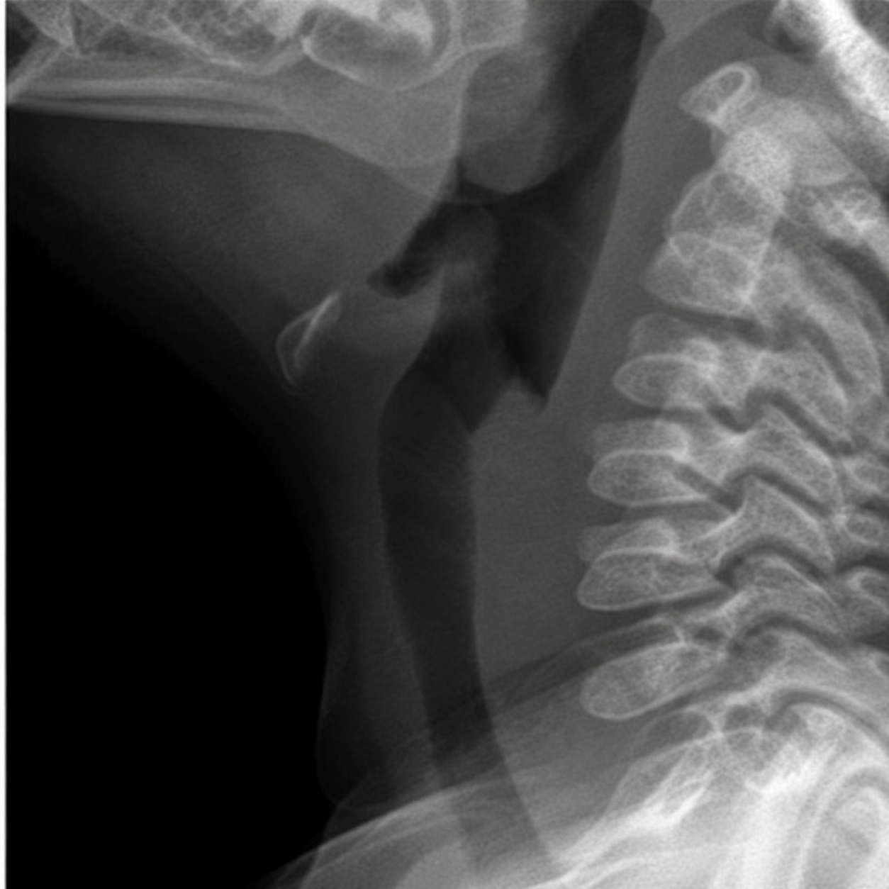
A lateral neck radiograph may demonstrate widening of the prevertebral soft tissues. Interpretation depends heavily on proper positioning and technique, and an abnormal radiograph does not reliably distinguish cellulitis or phlegmon from a mature abscess.
CT neck with IV contrast
In an airway-stable child, contrast-enhanced CT is commonly used to define the location and extent of deep neck infection and identify features concerning for a fluid collection or abscess.
Normal lateral neck
Retropharyngeal abscess
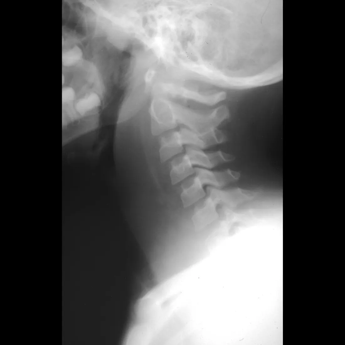
Compare the prevertebral soft tissues
Look anterior to the cervical vertebral bodies. Retropharyngeal infection can produce marked widening of the prevertebral soft tissues and anterior displacement of the airway.
Pediatric prevertebral soft-tissue landmarks
C2
Prevertebral soft tissue >7 mm is abnormal. At C1–C4, the soft-tissue thickness should generally remain less than half the AP width of the adjacent vertebral body.
C6
Prevertebral soft tissue >14 mm is abnormal. Below C4, the normal soft-tissue shadow may be thicker because of the esophagus.
C2 and C6 are commonly used reference levels. Assess the entire prevertebral contour and radiographic technique rather than relying on a single measurement.
Technique matters: Neck flexion and expiratory imaging can falsely increase apparent prevertebral soft-tissue thickness. These images are from different patients and are not displayed at identical radiographic scale; compare the overall contour rather than making direct measurements between images.
Contrast-enhanced CT of the neck
CT better defines the location and extent of deep neck infection when complications or a drainable collection are suspected.
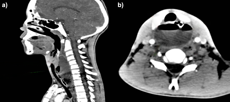
A, sagittal: The retropharyngeal collection extends along the cervical spine posterior to the airway. B, axial: Contrast-enhanced CT better defines the collection, surrounding deep neck spaces, and associated mass effect.
CT images adapted from Alzayed S, AlOlaywi A. Management of a Massive Retropharyngeal Abscess Through an External Cervical Approach: A Case Report. Cureus. 2025;17(12):e98740. CC BY 4.0. Original figure modified to remove source annotations; PediAtlas annotations added.
Reading the CT
Think in terms of a spectrum
Cellulitis
Inflammatory soft-tissue thickening and edema without a well-organized fluid collection.
Phlegmon
More organized inflammatory change may appear mass-like or heterogeneous but may still lack a clearly drainable purulent cavity.
Abscess
A discrete low-attenuation collection, particularly with peripheral enhancement, increases suspicion for a mature abscess.
Imaging Pitfall
CT cannot perfectly tell you whether pus will be found
Contrast-enhanced CT is very useful for defining deep neck infection, but inflammatory phlegmon and developing abscess can overlap in appearance. Rim enhancement or a fluid-appearing collection should therefore be interpreted alongside the child's clinical course, airway status, examination, and ENT assessment rather than treated as an automatic indication for operative drainage.
Clinical Reasoning
Imaging answers an anatomic question—not the entire management question
CT helps answer where the infection is, how extensive it is, whether adjacent deep neck spaces are involved, and whether a collection is present. The decision to continue medical therapy or proceed to drainage depends on those findings together with airway compromise, severity, abscess characteristics, and response to treatment.
Management
Airway stability determines the first move
Management of retropharyngeal infection combines early airway assessment, IV antibiotics, ENT involvement, and selective surgical drainage. Not every radiographic abscess requires immediate surgery.
Airway compromise
Respiratory distress, stridor, inability to handle secretions, rapidly progressive swelling, or other evidence of impending obstruction requires urgent ENT and airway involvement. Stabilization takes priority over additional diagnostic testing.
Airway stable
Begin IV antibiotics, involve ENT early, provide supportive care, and closely reassess the clinical response. Many stable children can initially be managed medically rather than proceeding directly to operative drainage.
Airway Safety
A difficult airway should be anticipated
Deep neck infection can distort upper-airway anatomy and worsen rapidly. When airway intervention is required, coordinate with experienced airway personnel and ENT whenever possible rather than approaching the airway as a routine intubation.
Empiric Antibiotic Therapy
Cover the expected polymicrobial flora
Initial therapy should cover common upper respiratory and oral organisms, including streptococci, Staphylococcus aureus, and anaerobic organisms.
Common initial options
- • Ampicillin-sulbactam
- • Clindamycin
Selection should reflect local susceptibility patterns, allergy history, illness severity, and institutional guidance.
When to broaden for MRSA
Additional MRSA-active therapy, such as vancomycin, may be appropriate in severe disease, high-risk patients, or when the child fails to improve on initial therapy.
Medical Therapy vs Drainage
The response over time matters
Initial medical management
In a clinically stable child without airway compromise, IV antibiotics with close observation and ENT involvement can be appropriate initially. Reassess symptoms, examination, airway status, fever curve, and overall trajectory over the next 24–48 hours.
Consider operative drainage
- • Airway compromise or respiratory distress
- • Clinical deterioration despite medical therapy
- • Failure to improve after an adequate antibiotic trial
- • Large or well-organized drainable collection
- • Complications or extension into adjacent spaces
Management Pearl
A collection on CT does not automatically equal surgery
In an airway-stable child, the decision to drain should integrate imaging with the clinical examination and response to IV antibiotics. Imaging can suggest abscess formation, but it does not perfectly predict whether operative exploration will reveal drainable purulence.
Reassessment
Watch the child—not just the scan
Improving fever, neck mobility, swallowing, oral intake, pain, and overall appearance support continued medical management. Worsening airway symptoms, persistent systemic illness, or failure of local symptoms to improve should prompt reconsideration of drainage and repeat ENT assessment.
Complications
A deep neck infection does not have to stay in the neck
The retropharyngeal space sits beside the upper airway and communicates with other deep cervical fascial planes. Progressive infection can therefore threaten the airway, spread into neighboring spaces, or descend into the chest.
Upper-airway obstruction
Expanding inflammation, edema, and abscess can displace the posterior pharyngeal wall anteriorly and progressively narrow the upper airway. Respiratory distress or stridor represents an immediate escalation in severity.
Descending mediastinitis
Infection can spread inferiorly through communicating deep cervical fascial planes toward the mediastinum. Descending mediastinal infection is a severe complication that can produce extensive thoracic infection and systemic illness.
Sepsis and bacteremia
Progressive or inadequately controlled infection can extend beyond the original deep neck space and produce bacteremia, sepsis, and systemic deterioration.
Adjacent deep-space spread
Infection may extend into neighboring cervical spaces rather than remaining confined to the retropharyngeal compartment, increasing disease extent and potentially altering the drainage approach.
Vascular complications
Deep cervical infection can involve nearby major vessels, producing rare but serious complications such as internal jugular vein thrombosis, carotid involvement, or hemorrhage.
Aspiration or pulmonary infection
Purulent material and impaired handling of secretions can contribute to aspiration and lower respiratory infection, particularly in children with advanced disease.
Anatomy → Complication
This is why the sagittal anatomy matters
The danger of retropharyngeal infection is determined partly by where it lives. The deep cervical fascial planes create potential pathways for infection to extend inferiorly from the neck toward the mediastinum. A child whose disease is spreading beyond the original space can therefore become critically ill even without dramatic findings on the initial oropharyngeal examination.
Clinical Warning
A worsening trajectory should trigger a search for complications
New respiratory symptoms, chest symptoms, increasing neck swelling, persistent fever or toxicity, hemodynamic instability, or failure to improve despite appropriate therapy should prompt reassessment for uncontrolled or extending infection.
Disposition
Admission is about the airway and the trajectory
Children with retropharyngeal abscess generally require hospitalization for IV antibiotics, airway observation, ENT evaluation, and serial reassessment.
Hospital admission
Admit for IV antibiotic therapy, hydration and analgesia as needed, ENT involvement, and repeated assessment of airway status, swallowing, neck mobility, fever, and overall clinical trajectory.
Higher-acuity care
Respiratory distress, progressive airway symptoms, inability to manage secretions, sepsis, rapidly worsening disease, or need for advanced airway management warrants escalation to a closely monitored or intensive care setting.
Improving child
Clinical improvement should include a stable airway, improving fever and pain, better neck mobility and swallowing, and the ability to maintain hydration as therapy transitions toward an outpatient regimen.
Discharge Readiness
Discharge follows clinical improvement—not simply a better scan
A child may be considered for discharge once the airway remains stable, symptoms and examination are clearly improving, oral intake and hydration are adequate, and an appropriate oral antibiotic plan and follow-up strategy are established.
Escalation
Reconsider the plan when the trajectory is wrong
New or worsening stridor, respiratory distress, drooling or inability to handle secretions, increasing neck swelling or stiffness, persistent systemic illness, or failure to improve with IV antibiotics should prompt urgent reassessment and reconsideration of airway intervention, drainage, or additional evaluation for complications.
Clinical Pearls
The high-yield takeaways
Retropharyngeal abscess is a disease of anatomy, age, and trajectory. Recognizing the localization early can identify deep neck infection before airway compromise develops.
Think RPA in the young child with fever + torticollis
The throat examination may look surprisingly unimpressive
Airway stability comes before imaging
CT defines anatomy better than it predicts pus
Not every radiographic abscess needs immediate drainage
Know where the deep neck spaces can lead
One-Line Summary
Fever + torticollis + dysphagia in a young child = think deep neck
Localize first, assess the airway, define the anatomy when it is safe to do so, begin appropriate antimicrobial therapy, and follow the child's clinical trajectory when deciding whether drainage is necessary.
Knowledge Check
Can you manage the child—not just recognize the abscess?
Work through five clinical decisions that integrate anatomy, presentation, airway assessment, imaging, and management.
Question 1
A 3-year-old develops fever, poor oral intake, drooling, and progressive refusal to extend his neck after several days of upper respiratory symptoms. Which finding most strongly localizes the infection to a deep neck space?
Anki Deck
Retropharyngeal Abscess Anki Deck
Reinforce the highest-yield concepts from this module with 16 spaced-repetition cards.
Requires Anki. Deck content is intended for educational review and should be used alongside the full PediAtlas module.
Evidence Base
Clinical References
Pediatric-focused review covering clinical presentation, deep-neck localization, radiographic evaluation, microbiology, complications, and management of retropharyngeal abscess.
Pediatric study evaluating factors associated with successful nonsurgical treatment of retropharyngeal and parapharyngeal infections and supporting selective medical management in appropriate children.
Study evaluating the ability of CT findings to distinguish cellulitis from abscess in pediatric deep neck infection, highlighting the limitations of imaging alone in predicting drainable purulence.
National pediatric study describing epidemiology and management trends, including the substantial proportion of retropharyngeal abscesses managed without surgical drainage.
Clinical review of retropharyngeal anatomy, presentation, imaging, antimicrobial therapy, airway concerns, complications, and operative management.
Secondary Educational Sources
Nelson Textbook of Pediatrics and Ferri's Clinical Advisor were used as additional textbook references for deep-neck anatomy, pathophysiology, microbiology, clinical presentation, imaging, antimicrobial therapy, complications, and operative decision-making.
Educational Disclaimer
PediAtlas is intended for medical education and should not replace institutional guidelines, local protocols, patient-specific assessment, or clinical judgment.