Peritonsillar Space & Oropharynx
Peritonsillar Abscess
A localized collection of pus in the peritonsillar space, most often affecting older children and adolescents and characterized by unilateral oropharyngeal swelling, odynophagia, muffled voice, and trismus.
Clinical Vignette
When pharyngitis becomes strikingly unilateral
Recognize the progression from tonsillar infection to a focal collection in the peritonsillar space.
A 15-year-old presents with several days of worsening sore throat and fever after initially developing symptoms consistent with acute pharyngotonsillitis.
Over the past 24 hours, the pain has become dramatically worse on the right. Swallowing is increasingly painful, oral intake has fallen, and the patient now speaks with a muffled voice.
Examination is difficult because the patient can only partially open the mouth because of trismus. The right peritonsillar region is prominently swollen, and the uvula is displaced away from the affected side.
The patient is uncomfortable but currently breathing without stridor or significant respiratory distress.
Your clinical reasoning task
Which findings suggest that this is no longer uncomplicated pharyngotonsillitis, and where has the infection localized?
Anatomy & Localization
Localize the infection to the peritonsillar space
The anatomy explains the asymmetric examination: PTA develops immediately outside the tonsillar capsule rather than within the tonsil itself.
PTA Anatomy & Localization
Compare what the disease looks like in the oropharynx with where the infection is located in axial anatomy.
Anterior Oropharyngeal View
What you see on physical examination
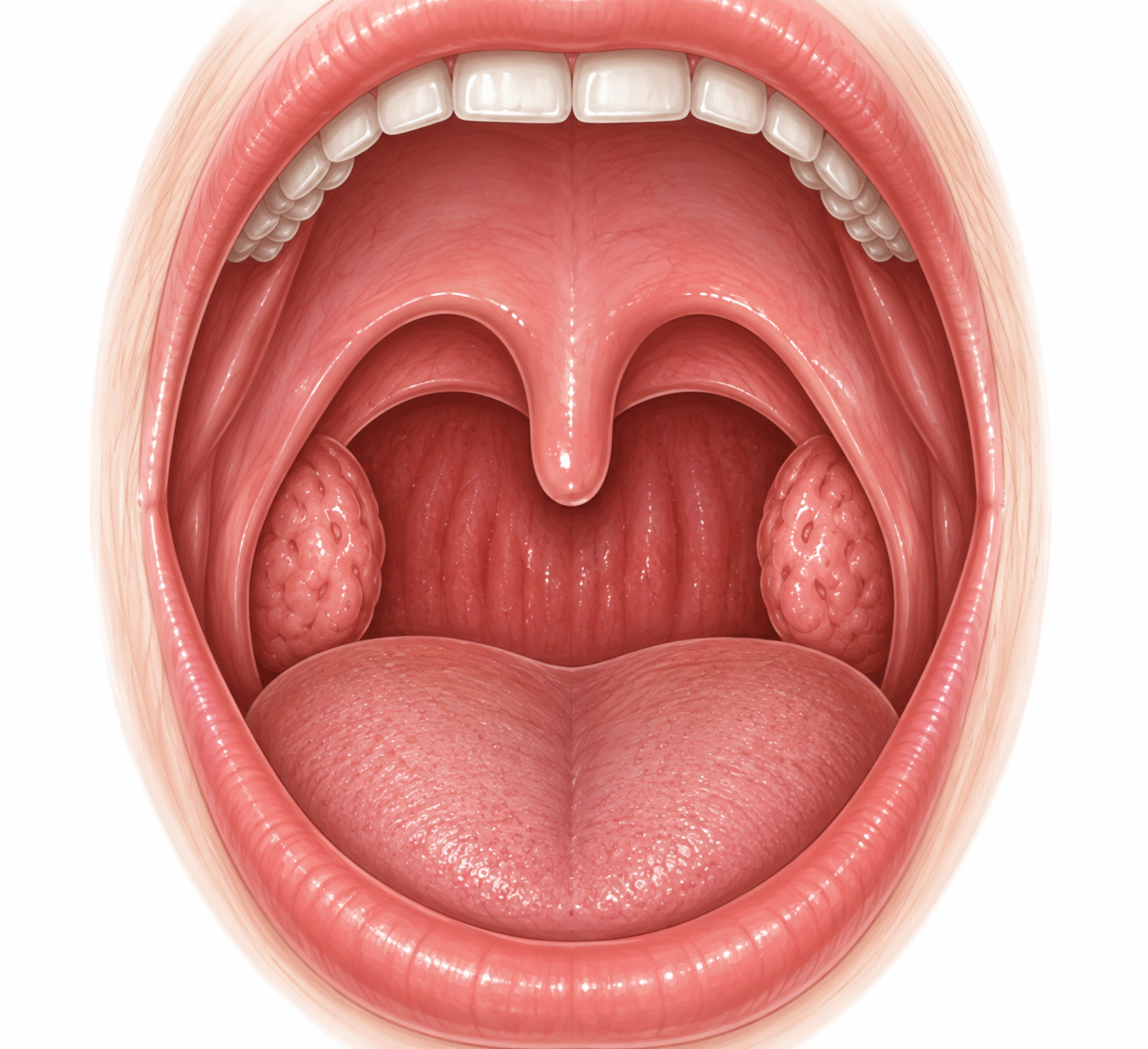
Normal landmarks
Axial View
Where the infection is anatomically
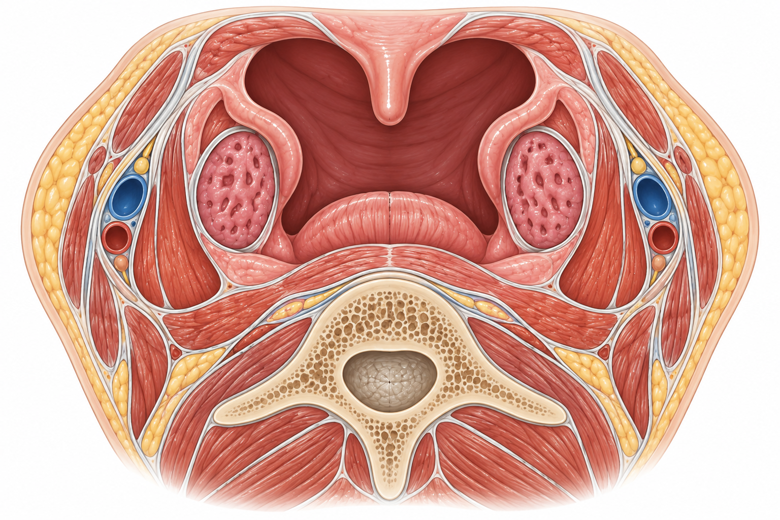
Normal axial landmarks
Axial orientation: anterior is at the top of the image and posterior is at the bottom.
Start with the palatine tonsil
- • The palatine tonsils sit along the lateral walls of the oropharynx.
- • Each tonsil lies between the anterior tonsillar pillar (palatoglossal arch) and posterior tonsillar pillar (palatopharyngeal arch).
- • A fibrous capsule separates the lateral surface of the tonsil from the adjacent pharyngeal musculature.
Then move just outside the capsule
- • The clinically important peritonsillar tissue lies between the tonsillar capsule and the pharyngeal constrictor musculature.
- • PTA represents a localized collection of pus in this peritonsillar region.
- • The collection is usually unilateral and commonly develops near the superior pole of the tonsil.
Anatomy Framework
Build the peritonsillar space from medial to lateral
Anterior boundary
- • The palatoglossal arch forms the anterior tonsillar pillar.
- • Inflammation may produce visible swelling and erythema of the anterior pillar and adjacent soft palate.
Posterior boundary
- • The palatopharyngeal arch forms the posterior tonsillar pillar.
- • Together, the tonsillar pillars frame the palatine tonsil within the oropharynx.
Lateral relationship
- • The superior pharyngeal constrictor lies lateral to the tonsillar capsule.
- • Spread beyond the peritonsillar region raises concern for deeper neck-space involvement.
Why does the uvula deviate?
A unilateral peritonsillar collection expands into the oropharynx, producing asymmetric soft-palate and tonsillar-region swelling. The affected tonsil may be displaced medially and inferiorly while the uvula is pushed toward the opposite side.
Why does the patient develop trismus?
Inflammation in the nearby masticator region can provoke painful reflex spasm of the muscles involved in opening the mouth. Clinically, trismus is an important clue that helps distinguish PTA from routine uncomplicated pharyngitis.
Anatomic Danger
Disease may extend beyond the peritonsillar space
Pathophysiology
From inflammation to a focal collection of pus
PTA is usually understood as suppurative infection of the peritonsillar tissues, often following acute tonsillar or pharyngeal infection.
Oropharyngeal infection
Many patients have preceding acute pharyngitis or tonsillitis, producing inflammation around the palatine tonsil and adjacent soft tissues.
Peritonsillar inflammation
Infection and inflammation involve the tissues immediately outside the tonsillar capsule. At this stage, peritonsillar cellulitis may exist without a discrete collection of pus.
Suppuration
Progressive infection, tissue necrosis, and suppuration produce a localized collection of pus in the peritonsillar region.
Local mass effect
The unilateral collection produces asymmetric soft-palate and peritonsillar swelling, medial displacement of the tonsillar region, and contralateral deviation of the uvula.
Trismus develops
Inflammation and reflex spasm of nearby muscles of mastication limit mouth opening and can make direct visualization of the peritonsillar region difficult.
Pathogenesis Nuance
Not every PTA necessarily follows a simple tonsillitis → cellulitis → abscess sequence
Clinical Translation
The pathophysiology predicts the bedside findings
History & Physical
Look for a strikingly unilateral oropharyngeal process
PTA usually presents with severe unilateral throat symptoms plus asymmetric peritonsillar findings. Trismus, muffled voice, and displacement of the tonsil and uvula help localize the infection beyond uncomplicated pharyngotonsillitis.
1. Pharyngeal symptoms
Fever and sore throat often precede abscess formation. As the infection localizes, throat pain typically becomes severe and predominantly unilateral, with increasing odynophagia and difficulty maintaining oral intake.
2. Local mass effect
Progressive peritonsillar swelling produces increasing oropharyngeal asymmetry. Patients may develop a muffled "hot potato" voice, difficulty swallowing, and ipsilateral referred ear pain.
3. Trismus & airway concern
Inflammation involving nearby muscles of mastication can produce trismus. Drooling, inability to handle secretions, respiratory distress, or rapidly progressive swelling should raise concern for more severe disease and possible airway compromise.
History clues
- • Severe sore throat, often predominantly unilateral
- • Fever
- • Odynophagia or dysphagia
- • Decreased oral intake
- • Muffled or "hot potato" voice
- • Ipsilateral referred otalgia
- • Drooling or difficulty handling secretions
Physical examination
- • Unilateral peritonsillar and soft-palate swelling
- • Medial displacement of the affected tonsil
- • Uvular deviation away from the affected side
- • Trismus limiting mouth opening
- • Tonsillar and pharyngeal erythema
- • Tender cervical lymphadenopathy
- • Assess airway and ability to handle secretions
Bedside Localization
Trismus plus unilateral peritonsillar swelling is a powerful clue
Uncomplicated tonsillitis can cause substantial throat pain and tonsillar inflammation, but marked asymmetry, peritonsillar or soft-palate bulging, and trismus should raise concern that infection has extended beyond the tonsil into the peritonsillar tissues.
Classic Finding
Uvular deviation supports the diagnosis—but do not require it
Expansion of a unilateral peritonsillar collection can displace the affected tonsillar region medially and push the uvula toward the opposite side. This is a classic examination finding, but its absence should not by itself exclude PTA when the remainder of the clinical picture is convincing.
Airway Check
Before focusing on the throat, make sure the patient can protect the airway
Assess work of breathing, voice, secretion handling, and the ability to swallow. Respiratory distress, progressive obstruction, or inability to manage secretions should prompt urgent escalation rather than an extended routine oropharyngeal examination.
Differential Diagnosis
Is this a PTA—or another cause of severe unilateral throat symptoms?
Severe throat pain, dysphagia, abnormal voice, and tonsillar inflammation overlap across several infections. Asymmetry, trismus, neck findings, and the location of swelling help narrow the diagnosis.
Peritonsillar cellulitis
The closest mimic of PTA. Peritonsillar tissues are inflamed and edematous, but there is no discrete purulent collection. Examination findings can overlap substantially with PTA, making the distinction difficult on examination alone.
Acute tonsillitis / pharyngitis
Can cause fever, severe throat pain, tonsillar erythema, enlargement, and exudate. Marked unilateral peritonsillar bulging, significant trismus, and displacement of surrounding structures favor PTA.
Retropharyngeal abscess
More often produces deep neck symptoms such as painful limitation of neck movement, stiffness, or torticollis, particularly in younger children. The infection is posterior to the pharynx rather than centered around the tonsillar capsule.
Parapharyngeal abscess
A lateral deep-neck infection that may cause fever, neck pain or swelling, torticollis, dysphagia, and trismus. Cross-sectional imaging may be needed when examination suggests infection extending beyond the peritonsillar region.
Epiglottitis
Severe dysphagia, drooling, muffled voice, respiratory distress, tripod positioning, or reluctance to lie flat favor supraglottic disease. Focal unilateral peritonsillar swelling and uvular displacement instead favor PTA.
Infectious mononucleosis
Can produce marked tonsillar enlargement, exudative pharyngitis, fever, and cervical lymphadenopathy. Tonsillar disease is often bilateral, while focal unilateral peritonsillar bulging and trismus should raise concern for PTA or a superimposed abscess.
Highest-Yield Distinction
Peritonsillar cellulitis and PTA lie on the same clinical spectrum
Peritonsillar cellulitis
Inflammation and edema involve the peritonsillar tissues, but a discrete purulent collection has not formed.
Peritonsillar abscess
Suppuration has produced a localized collection of pus in the peritonsillar region, potentially requiring drainage in addition to antimicrobial therapy.
Diagnostic Pearl
The examination may strongly suggest PTA without proving that pus is present
Trismus, unilateral peritonsillar swelling, tonsillar displacement, and uvular deviation can make PTA clinically likely, but peritonsillar cellulitis may produce overlapping findings. When the distinction remains uncertain and would change management, ultrasonography can help identify a discrete collection.
Deep Extension
Neck findings should make you reconsider whether the infection is still confined to the peritonsillar region
Prominent neck swelling, torticollis, painful limitation of neck movement, systemic deterioration, or other findings that seem disproportionate to a localized PTA should raise concern for parapharyngeal, retropharyngeal, or other deep-neck extension.
Diagnostic Workup
Most PTAs are diagnosed at the bedside
The classic combination of unilateral peritonsillar swelling, severe throat pain, trismus, muffled voice, and uvular displacement can make PTA a clinical diagnosis. Imaging is most useful when the diagnosis is uncertain or infection may extend beyond the peritonsillar space.
First Question
Does this look like a localized PTA—or something deeper?
Before ordering tests, assess the airway and determine whether the examination fits a localized peritonsillar process. Respiratory distress, inability to handle secretions, prominent neck swelling, torticollis, or other findings suggesting deep-neck extension should change the diagnostic approach and urgency.
Clinical examination
PTA is often diagnosed clinically. Look for unilateral peritonsillar and soft-palate swelling, medial displacement of the affected tonsil, trismus, muffled voice, and deviation of the uvula away from the affected side.
Ultrasound
Intraoral or transcervical ultrasonography can help distinguish peritonsillar cellulitis from a discrete abscess when the examination is equivocal. Ultrasound can also help define the location of a collection before drainage.
CT neck with IV contrast
CT is generally reserved for an atypical presentation, an examination limited by severe trismus, diagnostic uncertainty that cannot be resolved clinically or with ultrasound, or concern that infection has extended into adjacent deep-neck spaces.
Ultrasound of a Peritonsillar Abscess
Ultrasound can help distinguish a drainable fluid collection from peritonsillar cellulitis when the physical examination is equivocal.
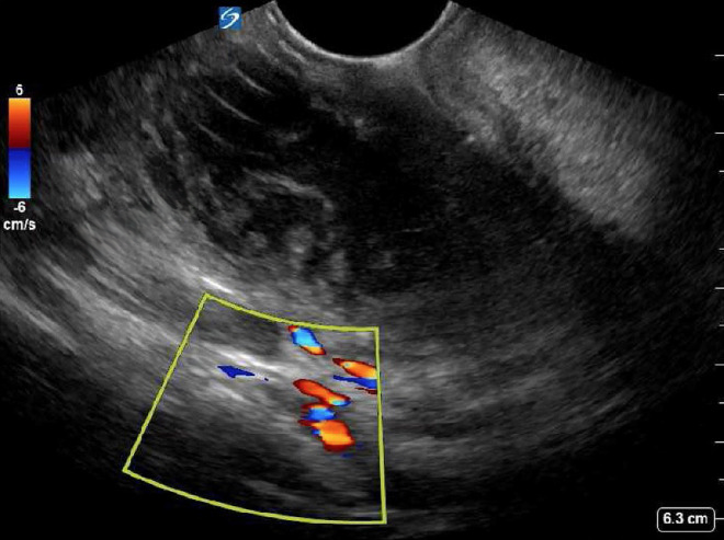
What to look for: A discrete hypoechoic or complex fluid collection supports abscess formation. Color Doppler can help demonstrate vascular structures adjacent to the collection and may assist procedural planning.
Source: Floyd J, Dahl B, Whited M, Kurzweil A. Peritonsillar Abscess and Post-aspiration Bleed Identified with Point-of-care Ultrasound Using Endocavitary Probe: A Case Report. CPC-EM Capsule. 2024;8(2):129–132. CC BY 4.0. Image cropped and annotated by PediAtlas.
A Practical Imaging Ladder
Escalate imaging according to the clinical question
1. Classic examination
A convincing unilateral peritonsillar examination may establish the diagnosis without routine cross-sectional imaging.
2. Cellulitis or abscess?
When the diagnosis is uncertain, ultrasound can look for a discrete fluid collection without exposing the patient to ionizing radiation.
3. Disease beyond the tonsil?
When the presentation suggests parapharyngeal, retropharyngeal, or other deep-neck extension, contrast-enhanced CT can define the involved spaces and surrounding structures.
Laboratory studies
Routine laboratory testing is not required to establish PTA in every patient. CBC, inflammatory markers, blood cultures, or other studies may be useful when the patient appears systemically ill, has significant dehydration, or there is concern for sepsis or a more extensive infection.
Microbiology
PTA is typically polymicrobial, involving organisms from the oropharyngeal flora. When purulent material is obtained during drainage, culture may be considered, particularly in severe, recurrent, unusual, or treatment-refractory infection.
Imaging Pearl
Ultrasound answers a different question than CT
Ultrasound is particularly useful when the bedside question is whether a discrete peritonsillar collection exists. CT becomes more valuable when the question changes to how far the infection extends and whether adjacent deep-neck spaces or critical structures are involved.
Important Pitfall
Do not make CT a routine prerequisite for drainage
A patient with a classic localized PTA does not necessarily need CT simply to confirm the diagnosis before treatment. Unnecessary cross-sectional imaging adds radiation and may delay definitive management without answering a clinically important question.
Return to the Anatomy
Deep Extension is the finding that changes the imaging strategy
The anatomy figure's progression from localized PTA to lateral deep-neck extension is also a diagnostic framework. Once symptoms or examination findings suggest that infection is no longer confined to the peritonsillar region, imaging should define the deeper spaces and their relationship to the airway and major cervical structures.
Management
Treat the infection—and decide whether the collection needs drainage
Management combines supportive care, antimicrobial therapy, and source control when appropriate. Airway status, illness severity, ability to tolerate oral intake, abscess characteristics, and clinical response help determine the treatment approach.
1. Assess stability
Evaluate the airway, secretion handling, hydration, and overall appearance first. Respiratory distress, progressive obstruction, or inability to manage secretions requires urgent escalation and ENT involvement.
2. Control symptoms
Provide adequate analgesia and hydration. Severe odynophagia can substantially limit oral intake, and some patients require IV fluids until pain and swallowing improve.
3. Treat the infection
Begin antimicrobial therapy directed against the polymicrobial oropharyngeal flora associated with PTA. The route and regimen depend on illness severity, oral tolerance, allergy history, local susceptibility patterns, and institutional guidance.
Airway First
A large abscess is important—a threatened airway is more important
If the patient develops respiratory distress, progressive upper-airway obstruction, or inability to handle secretions, airway stabilization and urgent specialty involvement take priority over routine imaging or bedside drainage planning.
Antimicrobial Therapy
Cover streptococci and oral anaerobes
PTA is usually polymicrobial. Antibiotic selection should provide coverage for common oropharyngeal pathogens, including group A streptococci and anaerobic organisms.
Parenteral therapy
- • Ampicillin-sulbactam is a commonly used option.
- • Other regimens may be selected according to allergy history, local susceptibility patterns, and illness severity.
- • Broader coverage may be appropriate in severe or complicated infection.
Oral therapy
- • Oral therapy is appropriate when the patient can reliably swallow and maintain hydration.
- • Amoxicillin-clavulanate is a commonly used oral option.
- • Antibiotic choice should follow local guidance and individual patient factors.
Source Control
Does this abscess need drainage?
Many established PTAs are managed with drainage plus antibiotics, but treatment should be individualized. Abscess size, severity, airway status, oral intake, age and cooperation, and response to therapy all influence the decision.
Selected medical management
Carefully selected patients with limited disease and reassuring airway status may improve with antimicrobial therapy and close reassessment without immediate drainage. The threshold for procedural management depends on the clinical course and local practice.
Drainage becomes more important
- • A well-formed or clinically significant abscess
- • Significant symptoms or inability to maintain hydration
- • Clinical deterioration
- • Failure to improve with appropriate medical therapy
- • Complicated or extending infection
Drainage Options
Source control can be achieved in several ways
Needle aspiration
Needle aspiration can both confirm the presence of purulence and provide therapeutic drainage in an appropriately selected and cooperative patient.
Incision & drainage
Incision and drainage provides direct evacuation of the collection and is a commonly used procedural approach for an established PTA.
Tonsillectomy
Immediate tonsillectomy is not required for every PTA but may be considered in selected patients based on recurrence, severity, airway considerations, or other indications for tonsillectomy.
Procedural Safety
Remember what lies lateral to the abscess
The carotid space and major cervical vascular structures lie lateral and posterolateral to the peritonsillar region. Drainage should respect this anatomy, and unusual anatomy or suspected deep extension should lower the threshold for specialty management and additional imaging rather than blind procedural exploration.
Reassessment
Treatment success should be visible at the bedside
Improvement should include decreasing pain and fever, better mouth opening, improved swallowing and oral intake, and reduction in peritonsillar swelling. Persistent or worsening symptoms should prompt reconsideration of an undrained collection, inadequate antimicrobial therapy, or extension into another deep-neck space.
Complications
Most PTAs remain localized—but extension can become dangerous
Complications arise from progressive local swelling, spread into adjacent deep-neck spaces, involvement of nearby vascular structures, aspiration, or systemic dissemination of infection.
Airway compromise
Progressive peritonsillar and pharyngeal swelling can narrow the upper airway. Respiratory distress, increasing obstruction, or inability to handle secretions represents an urgent escalation in severity.
Deep-neck extension
Infection may spread beyond the peritonsillar region into adjacent deep-neck spaces, including the parapharyngeal space. Extension changes both the risk profile and the approach to imaging and source control.
Vascular complications
Extension toward the lateral deep-neck spaces can place major cervical vessels at risk. Rare complications include internal jugular vein thrombosis and other infectious or vascular complications involving nearby structures.
Aspiration
Rupture or drainage of purulent material into the oropharynx, particularly when secretion handling is impaired, creates a risk of aspiration into the lower respiratory tract.
Sepsis
Local infection can progress to bacteremia and systemic illness. Persistent fever, toxicity, hemodynamic instability, or worsening clinical status should raise concern for uncontrolled or extending infection.
Recurrence
PTA can recur after successful treatment. Recurrence is one reason the history of previous PTA and other indications for tonsillectomy may influence longer-term ENT management.
Anatomy → Complication
The lateral boundary is what makes extension dangerous
A localized PTA begins immediately lateral to the tonsillar capsule, but infection that progresses beyond the pharyngeal musculature can enter deeper cervical spaces containing major neurovascular structures. This is why new neck swelling, torticollis, worsening systemic illness, or an atypical examination should trigger concern for disease beyond a simple localized PTA.
Clinical Warning
A changing examination matters more than the original diagnosis
Increasing neck swelling, worsening trismus, new respiratory symptoms, inability to handle secretions, persistent fever or toxicity, or failure to improve after appropriate therapy should prompt reassessment for inadequate source control or extension into another deep-neck space.
Recurrence
Successful drainage does not make future PTA impossible
Recurrent PTA occurs in a minority of pediatric patients. A history of recurrent abscesses or other established indications for tonsillectomy should prompt consideration of longer-term ENT management rather than treating each episode in isolation.
Disposition
Disposition depends on the airway, hydration, and response to treatment
Many patients with uncomplicated PTA can be managed without prolonged hospitalization once pain is controlled, oral intake is adequate, and the airway is reassuring. Admission is appropriate when those conditions are not met or the infection is complicated.
Consider discharge
- • Airway is stable
- • Able to handle secretions
- • Pain is adequately controlled
- • Able to tolerate oral fluids and medications
- • No evidence of deep-neck extension or systemic instability
- • Reliable follow-up and return precautions are established
Consider admission
- • Inability to maintain hydration or take oral medications
- • Poorly controlled pain or significant ongoing symptoms
- • Need for continued IV therapy or observation
- • Failure to improve after initial treatment
- • Significant comorbidity or concern for complicated infection
- • Uncertain ability to obtain appropriate follow-up
Higher-acuity care
- • Respiratory distress or progressive airway obstruction
- • Inability to handle secretions
- • Hemodynamic instability or sepsis
- • Rapidly progressive swelling
- • Significant deep-neck extension or other serious complication
- • Need for advanced airway management
Discharge Readiness
Make sure the patient can actually drink before sending them home
Improvement in the appearance of the throat alone is not enough. Before discharge, confirm that pain is controlled well enough for the patient to swallow, maintain hydration, and reliably take prescribed oral therapy.
Follow-Up
The trajectory still matters after treatment
Arrange appropriate clinical follow-up after discharge, particularly after drainage or when symptoms were substantial. Continued improvement in pain, trismus, swallowing, fever, and peritonsillar swelling should be expected rather than simply assuming that the procedure or first antibiotic dose completed treatment.
Return Precautions
Worsening airway or deep-neck symptoms require urgent reassessment
New or worsening difficulty breathing, inability to swallow or handle secretions, increasing neck swelling, worsening trismus, recurrent or persistent fever, progressive pain, or clinical deterioration should prompt urgent reassessment for recurrent collection, inadequate source control, or extension beyond the peritonsillar space.
Clinical Pearls
The high-yield takeaways
Peritonsillar abscess is a disease of localization. Recognize the unilateral examination, distinguish cellulitis from a true collection when necessary, and identify when infection is no longer confined to the peritonsillar space.
Severe unilateral findings should make you think beyond tonsillitis
Trismus is an important localization clue
PTA and peritonsillar cellulitis can look very similar
CT is for the deeper question
Treatment is antibiotics plus source control when needed
Know when the disease has outgrown the diagnosis
One-Line Summary
Unilateral throat pain + trismus + peritonsillar bulging = localize to the peritonsillar space
Assess the airway first, decide whether a discrete collection needs to be demonstrated, treat the polymicrobial infection, obtain source control when appropriate, and reconsider the anatomy if the patient is not improving as expected.
Knowledge Check
Can you manage the child—not just recognize the abscess?
Work through five clinical decisions that integrate anatomy, presentation, airway assessment, imaging, and management.
Question 1
A 15-year-old presents with fever, severe right-sided throat pain, muffled voice, and difficulty opening the mouth. Examination shows prominent right peritonsillar swelling with the uvula displaced to the left. Which diagnosis best explains this presentation?
Anki Deck
Peritonsillar Abscess Anki Deck
Reinforce the highest-yield concepts from this module with 16 spaced-repetition cards.
Requires Anki. Deck content is intended for educational review and should be used alongside the full PediAtlas module.
Evidence Base
Clinical References
Pediatric systematic review of peritonsillar abscess management, including antibiotics, drainage strategies, recurrence, and complications.
Educational Disclaimer
PediAtlas is intended for medical education and should not replace institutional guidelines, local protocols, patient-specific assessment, or clinical judgment.