Trachea & Subglottic Airway
Bacterial Tracheitis
Bacterial tracheitis is a potentially life-threatening bacterial infection of the trachea characterized by airway inflammation, thick mucopurulent secretions, and pseudomembranous debris that can produce severe upper-airway obstruction.
Clinical Vignette
When severe croup stops behaving like croup
Recognize the change in clinical trajectory that should raise concern for bacterial tracheitis.
A 5-year-old boy is brought to the emergency department after several days of rhinorrhea, sore throat, hoarseness, and a barking cough. His parents initially thought he had another viral respiratory illness.
Over the past 12 hours, however, he has developed a temperature of 39.6°C/103.3°F and rapidly worsening respiratory distress. His cough remains harsh and brassy, and he now has prominent stridor at rest with suprasternal and intercostal retractions.
He appears ill and fatigued but is able to lie flat. He is not drooling and does not appear to have difficulty swallowing his secretions.
He receives dexamethasone and nebulized epinephrine for presumed severe croup, but his stridor and work of breathing improve very little.
Your clinical reasoning task
Which features suggest that this is no longer uncomplicated viral croup, and what dangerous airway process should move to the top of your differential?
Anatomy & Localization
Bacterial tracheitis obstructs the airway from within the trachea
The disease centers on the tracheal mucosa, where inflammation, edema, and thick adherent secretions can progressively narrow the airway.
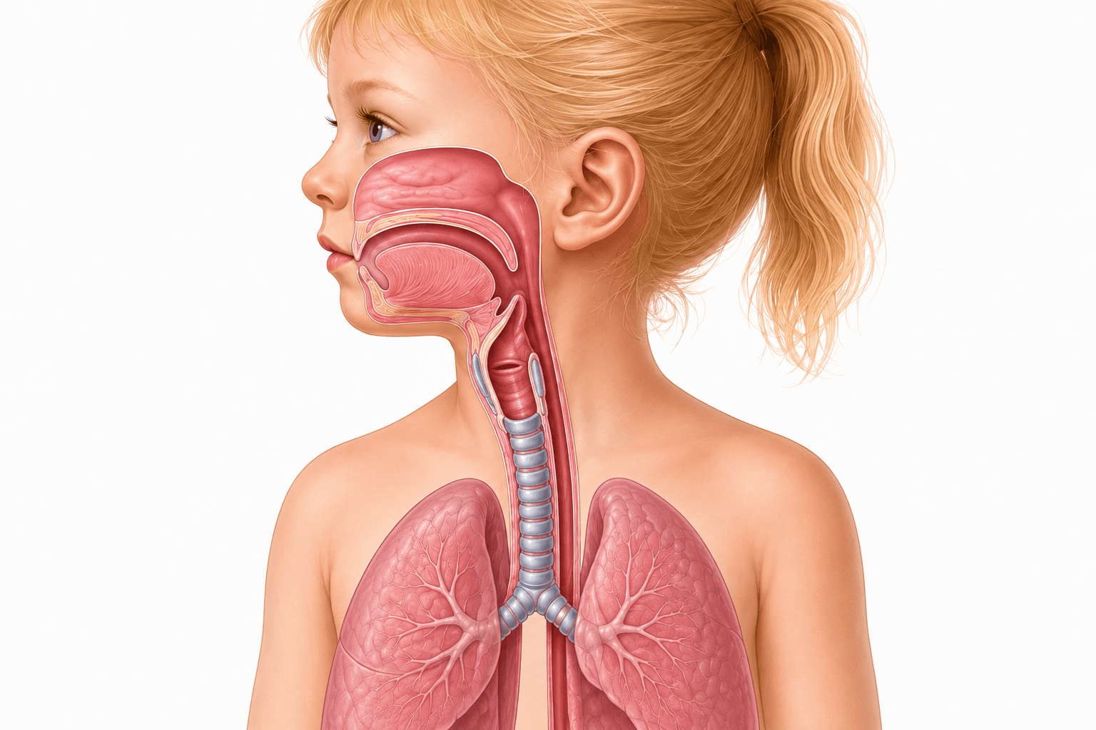
Trachea
The tracheal mucosa is the primary site of bacterial inflammation, where edema and thick secretions can progressively narrow the airway lumen.
Thick secretions & pseudomembranous debris
Mucopurulent secretions and sloughed inflamed mucosa can accumulate within the tracheal lumen and contribute directly to airway obstruction.
Tracheal airway
Bacterial tracheitis primarily involves the tracheal mucosa, often extending through the subglottic region. Inflammation causes mucosal edema while thick mucopurulent secretions and sloughed material accumulate within the airway lumen.
Why the obstruction is different from croup
Croup primarily narrows the subglottic airway through mucosal edema. Bacterial tracheitis can produce similar narrowing, but the airway is additionally obstructed by thick, tenacious secretions and pseudomembranous debris.
Mucosal edema
Bacterial inflammation thickens the tracheal mucosa and decreases the available airway diameter.
Purulent secretions
Copious, thick secretions can accumulate within the tracheal lumen and contribute directly to airway obstruction.
Pseudomembranous debris
Sloughed inflamed mucosa and adherent exudate may form irregular membranes that further narrow or intermittently obstruct airflow.
Localization Check
Same neighborhood as croup — different mechanism of obstruction
Both croup and bacterial tracheitis can produce stridor and subglottic narrowing. In bacterial tracheitis, however, infection extends into the trachea and creates thick purulent secretions and adherent debris that cannot be explained by edema alone.
Pathophysiology
Inflammation narrows the airway — secretions obstruct it from within
Bacterial tracheitis often follows viral injury to the tracheal mucosa, then adds bacterial inflammation and thick intraluminal debris to an already narrowed airway.
Viral injury primes the airway
A preceding viral respiratory infection can injure the tracheal mucosa and impair normal mucociliary defenses, creating a surface more susceptible to secondary bacterial infection.
Bacterial superinfection develops
Bacteria invade the injured tracheal mucosa and trigger intense local inflammation, producing erythema, edema, ulceration, and purulent inflammation.
Secretions and debris accumulate
Thick mucopurulent secretions combine with sloughed inflamed mucosa to form tenacious intraluminal material and, in some cases, adherent pseudomembranous debris.
Mechanical obstruction progresses
Mucosal edema narrows the airway from the outside inward while secretions and debris occupy the lumen from within, progressively increasing resistance to airflow.
Clinical Connection
Why doesn't this behave like uncomplicated croup?
Croup predominantly produces subglottic narrowing from mucosal edema. Bacterial tracheitis adds thick, tenacious secretions and inflammatory debris within the airway lumen. A child who appears increasingly toxic and continues to worsen despite appropriate croup therapy should prompt reconsideration of the diagnosis.
History & Physical
The child who was sick — then suddenly gets much sicker
The clinical course often provides the first clue: a viral or croup-like illness is followed by high fever, toxic appearance, and rapidly progressive upper-airway obstruction.
1. Viral prodrome
The illness often begins with several days of symptoms resembling a viral upper respiratory infection, including cough, rhinorrhea, sore throat, and fever.
2. Acute deterioration
The child then develops higher fever with worsening respiratory symptoms over hours, rather than following the expected improvement of an uncomplicated viral illness.
3. Toxic upper-airway disease
Stridor, hoarseness, respiratory distress, thick secretions, and a toxic or ill appearance should raise concern for bacterial tracheitis and impending airway obstruction.
History clues
- • Several-day viral or croup-like prodrome
- • Abrupt worsening rather than expected recovery
- • High fever
- • Increasing cough and respiratory distress
- • Poor or incomplete response to standard croup therapy
Physical examination
- • Toxic or markedly ill appearance
- • Inspiratory or biphasic stridor
- • Hoarseness or harsh cough
- • Increased work of breathing
- • Thick or mucopurulent airway secretions
- • Hypoxemia or altered mental status in severe obstruction
Special population
Children with tracheostomies or artificial airways
Tracheostomy- and ventilator-associated tracheitis may present more indolently than disease in a child with an intact airway. Look for a meaningful change from the child's respiratory baseline rather than relying on the classic croup-like presentation.
Secretions & suctioning
Increasing purulent secretions, changes in color, viscosity, or odor, or a new need for more frequent airway suctioning.
Respiratory support
Falling oxygen saturations or increasing ventilator requirements can signal clinically important airway infection.
We'll revisit this population during diagnostic workup, where distinguishing chronic airway colonization from active infection becomes especially important.
Differential Diagnosis
Stridor tells you where — the clinical pattern helps tell you why
Bacterial tracheitis overlaps with several causes of upper-airway obstruction. Fever, toxicity, secretion handling, illness trajectory, and response to therapy help separate the major mimics.
Epiglottitis
High fever and toxic appearance may overlap, but prominent dysphagia, drooling, muffled voice, tripod positioning, and refusal to lie down favor supraglottic disease.
Retropharyngeal / deep-neck infection
Fever with neck pain or stiffness, limited neck movement, dysphagia, drooling, muffled voice, or neck swelling should raise concern for a deep-neck space process.
Foreign body aspiration
Usually has abrupt onset after choking or eating, without a preceding febrile viral illness. Fever and progressive toxic appearance are less typical early in the course.
Anaphylaxis / angioedema
Rapid airway swelling may cause stridor but is usually accompanied by allergic features such as urticaria, lip or tongue swelling, gastrointestinal symptoms, hypotension, or a recent exposure.
Diphtheria
Consider in an incompletely immunized child or relevant exposure setting. Pharyngeal pseudomembrane and systemic toxicity distinguish it from typical bacterial tracheitis.
Highest-Yield Comparison
Croup vs bacterial tracheitis
Think uncomplicated croup
- • Barking cough and hoarseness dominate
- • Viral prodrome
- • Usually less toxic appearing
- • Lower or more modest fever is typical
- • Improves with standard croup therapy
Think bacterial tracheitis
- • Viral or croup-like prodrome followed by deterioration
- • High fever
- • Toxic or markedly ill appearance
- • Progressive stridor and respiratory distress
- • Thick or purulent airway secretions
- • Little or incomplete response to croup therapy
Diagnostic Workup
The airway comes before the workup
Bacterial tracheitis is primarily a clinical diagnosis. Testing should support the diagnosis without delaying stabilization of a threatened airway.
Threatened or unstable airway
Prioritize airway stabilization. Do not delay airway management for laboratory testing or imaging in a child with severe respiratory distress, hypoxemia, altered mental status, or rapidly progressive obstruction.
Stable enough for evaluation
When the airway is stable, focused laboratory studies and imaging can support the diagnosis, identify complications, and help distinguish bacterial tracheitis from competing diagnoses.
Laboratory studies
- • CBC may demonstrate leukocytosis or leukopenia but is nonspecific
- • CRP and ESR may be elevated but do not establish the diagnosis
- • Blood cultures have a low diagnostic yield
- • Obtain blood cultures when sepsis is suspected or the child is immunocompromised
Airway cultures
Tracheal secretions or exudates obtained during airway evaluation can be sent for Gram stain and bacterial culture. Results can identify the causative organism and guide narrowing of antimicrobial therapy.
Imaging findings
Expand for supportive radiographic findings. Imaging should only be obtained when the airway is sufficiently stable.
Imaging findings
Expand for supportive radiographic findings. Imaging should only be obtained when the airway is sufficiently stable.
Neck radiograph
AP or lateral neck imaging may demonstrate subglottic or tracheal narrowing. Irregularity or haziness of the tracheal air column may also occur. These findings are supportive rather than required for diagnosis.
Chest radiograph
Chest imaging is not diagnostic of bacterial tracheitis itself but can identify concurrent pneumonia or other lower-respiratory involvement.
Bronchoscopy
Bronchoscopy may demonstrate an inflamed tracheal mucosa with thick, adherent purulent secretions and intraluminal debris. It can be both diagnostic and therapeutic by allowing direct visualization, secretion sampling, and airway clearance.
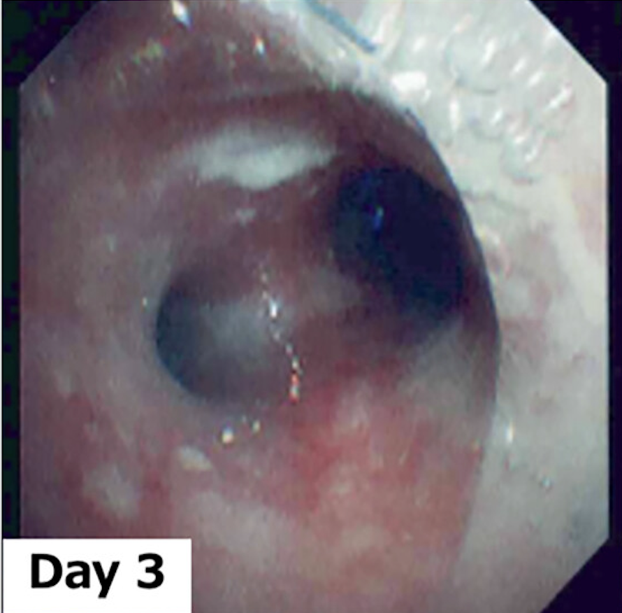
Bronchoscopic appearance of bacterial tracheitis
Bronchoscopy demonstrates inflamed tracheal mucosa with adherent purulent secretions near the tracheal bifurcation. Thick secretions and inflammatory debris can contribute to clinically significant airway obstruction.
Image: Kano K, et al. Pediatrics International. 2025. Licensed under CC BY 4.0.
Airway Safety
Never send a deteriorating child to imaging just to prove the diagnosis
Radiographs are adjuncts. If significant airway compromise is present, stabilization and definitive airway assessment take priority over obtaining a neck or chest image.
Direct airway evaluation
Bronchoscopy can diagnose and treat the obstruction
Direct visualization may demonstrate an erythematous, edematous trachea containing thick mucopurulent secretions or adherent pseudomembranous debris, while the epiglottis is normal or only mildly erythematous.
Diagnostic role
Directly demonstrates the abnormal tracheal mucosa and intraluminal secretions and allows specimens to be obtained for microbiologic testing.
Therapeutic role
Rigid bronchoscopy can permit suctioning and removal of thick secretions or adherent debris that are contributing directly to airway obstruction.
Direct visualization is not required in every clinically suspected case. The decision to perform bronchoscopy depends on disease severity and the need for airway intervention.
Tracheostomy Pearl
A positive tracheal culture does not automatically mean infection
Artificial airways are readily colonized by bacteria and can develop biofilms. Interpret tracheal cultures in the context of a meaningful clinical change rather than treating culture positivity alone.
Findings supporting active disease
- • New or increasingly purulent secretions
- • Increased suctioning requirement
- • Change in secretion color, viscosity, or odor
- • Fever or other systemic signs of illness
- • Worsening oxygenation or ventilatory requirements
Also evaluate for lower-airway disease
Isolated tracheitis is uncommon in children with artificial airways. Concurrent bronchial or pulmonary infection may be present, so chest imaging and the overall respiratory picture can help distinguish isolated tracheitis from pneumonia.
Management
Protect the airway, clear the obstruction, and treat the infection
Management is driven by the severity of airway obstruction. Children with progressive disease may require definitive airway control and removal of thick tracheal secretions in addition to antimicrobial therapy.
Assess airway severity
Closely monitor stridor, work of breathing, air entry, oxygenation, mental status, and the child's ability to sustain respiratory effort.
Secure a threatened airway
Progressive obstruction, hypoxemia, fatigue, altered mental status, or severe respiratory distress should prompt early definitive airway planning with experienced pediatric airway clinicians.
Clear secretions and debris
Thick tracheal secretions may require frequent suctioning or bronchoscopic removal because intraluminal material contributes directly to airway obstruction.
Start antimicrobial therapy
Begin empiric parenteral antibiotics directed against the major bacterial pathogens, including Staphylococcus aureus, then narrow therapy when culture and susceptibility data are available.
When airway intervention is needed
Do not wait for respiratory arrest before escalating airway management. Progressive bacterial tracheitis can deteriorate rapidly as edema and thick secretions increasingly narrow the tracheal lumen.
- • Severe or progressively worsening stridor
- • Increasing work of breathing or decreasing air entry
- • Hypoxemia or cyanosis
- • Fatigue or altered mental status
- • Inability to effectively clear thick secretions
- • Rapid clinical deterioration
Airway clearance & bronchoscopy
Thick secretions require aggressive airway clearance with suctioning and humidification. When secretions or adherent debris cannot be adequately cleared, bronchoscopy can directly remove obstructing material.
Bronchoscopy may therefore be therapeutic in addition to its diagnostic role, particularly in children with significant intraluminal obstruction.
Airway Pearl
Intubation secures the airway — it does not clear the disease
Thick secretions and sloughed inflammatory material can continue to obstruct the trachea or the endotracheal tube after intubation. Frequent suctioning, humidification, and repeated assessment of tube patency may still be necessary.
Empiric antimicrobial therapy
Initial parenteral therapy should cover the major bacterial causes of tracheitis, particularly Staphylococcus aureus, while also providing coverage for other common respiratory pathogens.
Typical empiric approach
A third-generation cephalosporin such as ceftriaxone or cefotaxime is commonly paired with antistaphylococcal therapy. MRSA-active coverage should be selected according to local susceptibility patterns and illness severity.
Once cultures return
Narrow antimicrobial therapy to the identified organism and susceptibility profile whenever possible.
Supportive care
- • Continuous cardiorespiratory and pulse-oximetry monitoring
- • Supplemental oxygen when indicated
- • IV fluids when oral intake is inadequate
- • Antipyretics and analgesia as appropriate
- • Frequent reassessment of respiratory status
Management considerations with a tracheostomy
Expand for additional considerations in children with artificial airways.
Management considerations with a tracheostomy
Expand for additional considerations in children with artificial airways.
Children with tracheostomies may require increased suctioning, airway clearance, humidification, and respiratory support during acute infection.
Because chronic colonization is common, antimicrobial decisions should integrate the child's clinical change, prior airway cultures, previous resistant organisms, and current microbiologic data rather than treating a positive culture in isolation.
Disposition
Admission is the rule — level of care follows the airway
Bacterial tracheitis can deteriorate rapidly. Disposition should reflect the degree of airway compromise, need for respiratory support, and ability to provide immediate airway intervention if the child worsens.
PICU / higher-acuity care
Children with significant or progressive airway obstruction require intensive monitoring and immediate access to experienced pediatric airway management.
- • Endotracheal intubation or mechanical ventilation
- • Progressive stridor or respiratory distress
- • Hypoxemia or increasing respiratory support
- • Fatigue, altered mental status, or decreasing air entry
- • Frequent suctioning for thick obstructing secretions
Stable but not intubated
Children who do not require intubation still need admission and close observation in a setting where deterioration can be recognized quickly and the airway can be secured if necessary.
If appropriate pediatric airway expertise is not available locally, transfer to a higher-level or tertiary-care center should be considered.
Extubation Readiness
Improvement means more than a better oxygen saturation
Extubation is individualized but should follow clear clinical improvement: fever and toxic appearance are resolving, tracheal secretions have substantially decreased, respiratory status has stabilized, and an air leak around the endotracheal tube supports improvement in airway edema.
After extubation
Continue close observation after extubation for recurrent obstruction while oxygen therapy, airway clearance, and antimicrobial treatment are continued as clinically indicated.
Discharge readiness
- • Stable airway without recurrent significant obstruction
- • No ongoing need for intensive respiratory support
- • Secretions manageable without intensive airway intervention
- • Adequate hydration and ability to tolerate enteral therapy
- • Appropriate antimicrobial plan established for completion
Expected Course
Most children recover completely with appropriate treatment
Improvement is generally expected over several days with effective airway management and antimicrobial therapy. Reported intubation duration is commonly a few days, although hospitalization may be longer depending on disease severity and associated lower-respiratory involvement.
Clinical Pearls
The high-yield takeaways
Keep these principles in mind when evaluating a child with suspected bacterial tracheitis.
Think bacterial tracheitis when croup stops behaving like croup
A viral or croup-like prodrome followed by high fever, toxic appearance, progressive respiratory distress, and poor response to standard croup therapy should prompt reconsideration of the diagnosis.
The obstruction is both edema and debris
Unlike uncomplicated croup, bacterial tracheitis can obstruct the airway with thick mucopurulent secretions and sloughed or pseudomembranous material in addition to mucosal edema.
Drooling should make you reconsider the localization
Children with bacterial tracheitis usually remain able to handle their oral secretions. Prominent drooling, dysphagia, muffled voice, or tripod positioning should increase concern for supraglottic disease such as epiglottitis.
The airway comes before the diagnostic workup
Imaging and laboratory testing are supportive. A child with progressive airway compromise should undergo stabilization and definitive airway assessment rather than being sent for testing simply to confirm the diagnosis.
Bronchoscopy can diagnose and treat
Direct airway visualization can demonstrate the inflamed trachea, obtain microbiologic specimens, and permit removal of thick secretions or adherent debris contributing to obstruction.
A positive trach culture is not the same as infection
Children with tracheostomies commonly have airway colonization. Interpret cultures alongside meaningful clinical changes such as new purulent secretions, increased suctioning, fever, or worsening respiratory support requirements.
Remember the Management Framework
Airway → clearance → antibiotics
Protect a threatened airway, remove obstructing secretions and debris when necessary, and begin empiric parenteral antimicrobial therapy with appropriate coverage for Staphylococcus aureus and other likely respiratory pathogens.
Progressive Quiz
Can you manage the deteriorating airway?
Follow the case as it evolves from a croup-like illness to progressive bacterial tracheal obstruction.
Question 1
A 4-year-old has had 3 days of rhinorrhea, cough, and low-grade fever. He now develops high fever, worsening stridor, increased work of breathing, and a toxic appearance. Nebulized epinephrine produces little improvement. Which diagnosis is most likely?
Spaced Repetition
Keep the high-yield concepts fresh
Review the core bacterial tracheitis concepts from this module with a focused Anki deck.
Anki Deck
Bacterial Tracheitis
Reinforce the highest-yield concepts from this module with 12 spaced-repetition cards.
Requires Anki. Deck content is intended for educational review and should be used alongside the full PediAtlas module.
Evidence Base
Clinical References
Pediatric-focused review of presentation, airway obstruction, diagnosis, antimicrobial therapy, disposition, and bacterial tracheitis in children with artificial airways.
Peer-reviewed clinical review addressing pathophysiology, differentiation from croup and epiglottitis, bronchoscopy, airway management, antimicrobial therapy, and prognosis.
Contemporary pediatric case series describing presenting features, viral coinfection, microbiology, PICU admission, intubation, and clinical outcomes.
Large pediatric database study highlighting important differences in presentation and management between children with and without preexisting tracheostomies.
Multicenter pediatric study examining diagnostic testing, antimicrobial practice variation, length of stay, and outcomes in children with tracheostomies hospitalized with bacterial respiratory infections.
Secondary Educational Sources
Nelson Textbook of Pediatrics and StatPearls were used as additional educational references for clinical organization, cross-checking, and consistency.
Educational Disclaimer
PediAtlas is intended for medical education and should not replace institutional guidelines, local protocols, patient-specific assessment, or clinical judgment.