Airway Protection & Swallowing Disorders
Pulmonary Aspiration
Pulmonary aspiration occurs when material from the oropharynx or stomach enters the lower respiratory tract. The clinical presentation ranges from acute chemical lung injury to bacterial pneumonia and chronic recurrent aspiration resulting in progressive lung disease.
Clinical vignette
An infant with coughing during feeds
A 5-month-old infant presents with recurrent coughing, choking, and oxygen desaturations during bottle feeds. Parents report frequent respiratory illnesses and poor weight gain. Chest radiographs demonstrate recurrent right lower lobe infiltrates. The clinical team becomes concerned for pulmonary aspiration caused by impaired swallowing.
Your first clinical task
Determine whether aspiration is acute or chronic, identify the source of aspirated material, evaluate swallowing safety, and recognize which aspiration syndrome best explains the child's presentation.
Anatomy & Localization
Aspiration begins when airway protection fails
During normal swallowing, multiple protective mechanisms prevent food, liquid, and gastric contents from entering the lower respiratory tract. Pulmonary aspiration occurs when these defenses fail, allowing material to pass through the larynx and into the tracheobronchial tree.
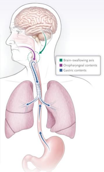
Normal Swallowing
Multiple protective mechanisms
- • Coordinated oral and pharyngeal swallow
- • Elevation of the larynx
- • Epiglottic inversion over the laryngeal inlet
- • Vocal cord closure
- • Upper esophageal sphincter relaxation
- • Effective cough clears minor aspiration events
Pulmonary Aspiration
Material enters the lower airway
- • Food or liquid passes below the vocal cords
- • Gastric contents may reflux into the airway
- • Poor cough fails to clear aspirated material
- • Repeated aspiration causes chronic inflammation
- • Infection develops when bacteria proliferate
- • Long-term injury may lead to bronchiectasis
Common causes of aspiration
Swallowing dysfunction
Neurologic disease, developmental delay, craniofacial abnormalities, and immature feeding coordination impair normal swallowing.
Reduced airway protection
Altered mental status, seizures, sedation, anesthesia, or intoxication reduce protective airway reflexes.
Gastroesophageal reflux
Refluxed gastric contents may reach the pharynx and be aspirated, particularly in children with impaired airway clearance.
Structural abnormalities
Laryngeal cleft, tracheoesophageal fistula, vocal cord paralysis, and other congenital lesions predispose to recurrent aspiration.
Pediatric Anatomy Pearl
Healthy children frequently experience small episodes of physiologic microaspiration during sleep that are rapidly cleared by normal cough and mucociliary function. Disease develops when aspiration is large-volume, recurrent, or occurs in children with impaired airway protection or ineffective clearance mechanisms.
Pathophysiology
Aspiration produces three clinically distinct syndromes
The pulmonary response depends on the type of material aspirated, the volume and frequency of aspiration, the bacterial burden, and the child's ability to protect and clear the airway. Distinguishing these syndromes helps determine whether treatment should emphasize supportive care, antibiotics, or prevention of recurrent aspiration.
Sterile chemical lung injury
Aspiration pneumonitis
Mechanism
Direct chemical injury after aspiration of acidic gastric contents or another irritating substance.
Typical timing
Abrupt symptoms developing within minutes to hours of a witnessed or strongly suspected aspiration event.
Clinical findings
Cough, tachypnea, hypoxemia, increased work of breathing, wheezing, or diffuse crackles. Fever and leukocytosis may occur from inflammation alone.
Imaging
Dependent or multifocal airspace opacities may appear, although early chest radiographs can be normal.
Treatment
Supportive respiratory care. Antibiotics are not routinely indicated unless a bacterial infection subsequently becomes likely.
Bacterial pulmonary infection
Aspiration pneumonia
Mechanism
Aspiration of bacteria-containing oropharyngeal or gastric material followed by infection of the pulmonary parenchyma.
Typical timing
Symptoms generally evolve over hours to days rather than immediately after a single aspiration event.
Clinical findings
Persistent or worsening fever, cough, respiratory distress, focal crackles, hypoxemia, and systemic signs of infection.
Imaging
Focal or multifocal infiltrates, often involving dependent lung segments based on the child's position during aspiration.
Treatment
Antibiotic therapy directed toward likely community or hospital pathogens, guided by the clinical setting and local recommendations.
Repeated or ongoing airway contamination
Chronic pulmonary aspiration
Mechanism
Repeated aspiration caused by swallowing dysfunction, impaired airway protection, reflux, structural abnormalities, or ineffective clearance.
Typical timing
A recurrent or progressive course over weeks to months, often without one dramatic aspiration event.
Clinical findings
Coughing or choking with feeds, wet respirations, recurrent pneumonia, chronic cough, wheezing, poor growth, or persistent oxygen requirement.
Imaging
Recurrent dependent opacities, atelectasis, airway wall thickening, bronchiectasis, or chronic inflammatory changes.
Treatment
Identify and address the source of aspiration, optimize feeding safety and airway clearance, and treat acute infections or chronic lung complications when present.
Clinical timeline pearl
Immediate respiratory deterioration after a clear aspiration event favors pneumonitis. Persistent or progressive infectious findings over the following days favor aspiration pneumonia. Recurrent feeding-associated symptoms and repeated focal lung disease suggest chronic pulmonary aspiration.
Histry and Phyiscal Exam
Look for acute events, feeding symptoms, and evidence of chronic lung injury
Aspiration may present as an abrupt respiratory event or as recurrent, subtle symptoms associated with feeding. A focused history and examination should identify the likely timing, source, severity, and underlying risk factors.
History
Ask how and when symptoms occur
Acute aspiration clues
- • Witnessed choking, gagging, or vomiting
- • Sudden cough or respiratory distress
- • Seizure or altered mental status
- • Recent anesthesia, sedation, or procedure
- • Abrupt oxygen desaturation
- • Symptoms beginning within hours of an event
Chronic aspiration clues
- • Coughing, choking, or gagging during feeds
- • Wet voice or wet respirations after feeding
- • Prolonged or stressful feeding sessions
- • Recurrent pneumonia or bronchiolitis-like illness
- • Chronic cough or persistent wheezing
- • Feeding refusal, fatigue, or poor weight gain
Physical examination
Examine breathing, feeding, and growth
Respiratory findings
- • Tachypnea or increased work of breathing
- • Hypoxemia or intermittent desaturation
- • Crackles, rhonchi, or wheezing
- • Decreased or asymmetric air entry
- • Persistent oxygen requirement
Feeding and general findings
- • Coughing or choking during observed feeds
- • Drooling or difficulty managing secretions
- • Wet vocal quality after swallowing
- • Poor coordination of suck, swallow, and breathing
- • Growth faltering or malnutrition
- • Digital clubbing in advanced chronic disease
Risk factors
Identify why airway protection may be impaired
- • Neurologic impairment, developmental delay, or neuromuscular weakness
- • Prematurity or immature suck-swallow-breathe coordination
- • Craniofacial abnormalities or congenital airway lesions
- • Laryngeal cleft, vocal cord paralysis, or tracheoesophageal fistula
- • Gastroesophageal reflux with impaired airway clearance
- • Tracheostomy, feeding tube dependence, or prolonged hospitalization
- • Sedating medications or recurrent episodes of altered consciousness
Red flags
Escalate when airway or respiratory stability is threatened
- • Apnea, cyanosis, or altered mental status
- • Severe hypoxemia or rapidly increasing oxygen need
- • Marked respiratory distress or exhaustion
- • Inability to protect the airway or manage secretions
- • Recurrent aspiration despite current feeding precautions
- • Failure to thrive with significant feeding intolerance
- • Recurrent pneumonia in the same dependent region
- • Concern for sepsis, abscess, or complicated pneumonia
Silent aspiration
The absence of coughing does not exclude aspiration
Children with neurologic impairment, reduced laryngeal sensation, or weak cough may aspirate without obvious choking. Recurrent respiratory disease, feeding difficulty, or unexplained oxygen desaturation may be the only clues.
Diagnostic workup
Choose testing based on the clinical question
No single study evaluates every cause or consequence of aspiration. Testing should determine whether aspiration is occurring, identify resulting lung injury, and clarify the underlying swallowing, airway, neurologic, or gastrointestinal disorder.
Clinical question 1
Is aspiration occurring during swallowing?
Clinical feeding evaluation
A speech-language pathologist assesses oral motor function, feeding coordination, positioning, secretion management, and symptoms during feeding.
Videofluoroscopic swallow study (VFSS)/Modified Barium Swallow Study (MBSS)
VFSS/MBSS evaluates the oral and pharyngeal phases of swallowing using contrast-containing liquids and foods. It can identify penetration, aspiration, timing abnormalities, and potentially safer consistencies or feeding strategies.
Flexible endoscopic evaluation of swallowing
FEES directly examines pharyngeal and laryngeal anatomy, secretion management, airway protection, and swallowing before and after the brief white-out period created by pharyngeal contraction.
Clinical question 2
Has acute pulmonary injury occurred?
Pulse oximetry and respiratory monitoring
Assess oxygenation, respiratory effort, and the need for escalating respiratory support.
Chest radiograph
Obtain when respiratory symptoms are significant, persistent, focal, or concerning for pneumonia or a complication. Early imaging may be normal despite a clinically important aspiration event.
Laboratory testing
CBC, inflammatory markers, blood gas, cultures, or metabolic testing should be guided by illness severity and concern for infection, respiratory failure, or an underlying acute event.
Clinical question 3
Is there recurrent or chronic lung injury?
Review prior imaging
Recurrent opacities, atelectasis, or disease repeatedly affecting dependent regions may suggest ongoing aspiration.
Chest CT
Consider when symptoms are persistent, complications are suspected, or detailed assessment for bronchiectasis, chronic atelectasis, airway disease, or structural lung abnormalities is needed.
Bronchoscopy
May be used to evaluate airway anatomy, retained material, infection, airway inflammation, or another diagnosis when noninvasive testing does not explain the clinical course.
Clinical question 4
Why is aspiration occurring?
- • Speech-language pathology: swallowing physiology and feeding safety
- • ENT: vocal cord dysfunction, laryngeal cleft, airway lesions, or impaired laryngeal sensation
- • Pulmonology: chronic lung injury, bronchoscopy, and airway clearance
- • Gastroenterology: esophageal disease, reflux evaluation, motility disorders, and nutrition
- • Neurology: impaired consciousness, neuromuscular weakness, seizures, or developmental disease
- • Aerodigestive team: coordinated evaluation when several systems contribute
Swallow imaging
Videofluoroscopic swallow study (VFSS)/Modified Barium Swallow Study (MBSS)

What to look for
Contrast passing below the level of the vocal folds indicates aspiration. Contrast entering the laryngeal vestibule but remaining above the vocal folds is termed penetration.
Chest imaging
Aspiration-related airspace disease

Imaging pearl
Aspiration often affects dependent lung regions, but the distribution varies with patient position and the timing of the event. Imaging findings are supportive rather than independently diagnostic of aspiration.
Important limitation
A single normal study does not exclude intermittent aspiration
Aspiration may vary by consistency, fatigue, positioning, illness, feeding technique, and level of alertness. Test results must be interpreted alongside the child's usual feeding history and respiratory course.
Management
Stabilize first, identify the syndrome, then prevent recurrence
Initial management focuses on airway protection and respiratory support. Subsequent treatment depends on whether the child has aspiration pneumonitis, aspiration pneumonia, or chronic pulmonary aspiration requiring long-term intervention.
Initial Management Algorithm
Unstable Patient
- • Airway protection
- • Supplemental oxygen
- • Suction secretions
- • Escalate respiratory support
- • Mechanical ventilation if needed
- • PICU consultation
Stable Patient
- • Focused history & physical
- • Determine aspiration syndrome
- • Obtain indicated studies
- • Identify underlying cause
Aspiration Pneumonitis
Supportive care. Routine antibiotics are not indicated.
Aspiration Pneumonia
Begin appropriate antibiotics plus supportive care.
Chronic Aspiration
Investigate the underlying cause and develop a long-term prevention strategy.
Supportive Care
- • Airway positioning and suctioning
- • Supplemental oxygen
- • Escalate respiratory support as needed
- • IV fluids when oral intake is inadequate
- • Bronchodilators only if bronchospasm is present
- • Continuous reassessment for clinical deterioration
Antibiotic Therapy
- • Not routinely recommended for isolated aspiration pneumonitis
- • Treat aspiration pneumonia when bacterial infection is suspected
- • Choose antibiotics based on community versus hospital acquisition
- • Adjust therapy using cultures and clinical response when available
Prevent Recurrence
- • Swallow therapy with Speech-Language Pathology
- • Feeding modifications when appropriate
- • Optimize positioning during feeds
- • Treat contributing neurologic or GI disorders
- • Multidisciplinary aerodigestive follow-up for complex patients
Clinical Pearl
The underlying cause is just as important as treating the acute event
Children with recurrent aspiration often require coordinated care involving Speech-Language Pathology, Pulmonology, Gastroenterology, Otolaryngology, Neurology, Nutrition, and Primary Care. Preventing future aspiration episodes is essential to avoid progressive chronic lung disease.
Disposition/Care setting & follow-up
Match the care plan to respiratory severity, feeding safety, and recurrence risk
Children with pulmonary aspiration may be managed in the outpatient setting, admitted for acute respiratory support or feeding evaluation, or require intensive care when airway protection and ventilation are compromised. Long-term follow-up should focus on preventing recurrent aspiration and chronic lung injury.
Outpatient care
Appropriate when the child is clinically stable
- • Stable oxygenation without significant respiratory distress
- • Safe feeding plan or reliable alternative nutrition plan
- • No evidence of progressive respiratory deterioration
- • Caregivers understand feeding precautions and warning signs
- • Timely primary care and subspecialty follow-up can be arranged
Inpatient care
Admit when monitoring or active treatment is required
- • Persistent oxygen requirement or moderate respiratory distress
- • Need for IV fluids, enteral support, or feeding restriction
- • Aspiration pneumonia requiring inpatient therapy
- • Need for swallow evaluation or feeding-plan development
- • Recurrent events requiring coordinated diagnostic evaluation
- • Inability to maintain hydration, nutrition, or secretion control
Intensive care
Escalate when airway protection or ventilation is threatened
- • Respiratory failure or rapidly worsening gas exchange
- • Need for noninvasive or invasive ventilatory support
- • Inability to protect the airway or manage secretions
- • Recurrent apnea, cyanosis, or altered mental status
- • Hemodynamic instability or concern for severe sepsis
Long-term follow-up
Follow-up should address both lung disease and the source of aspiration
Depending on the underlying disorder, children may require coordinated care involving Primary Care, Speech-Language Pathology, Pulmonology, Gastroenterology, Otolaryngology, Nutrition, Neurology, Surgery, and an aerodigestive program.
Monitor respiratory outcomes
- • Recurrent pneumonia or wheezing
- • Oxygen requirement
- • Airway-clearance needs
- • Development of bronchiectasis or chronic lung disease
Monitor feeding and growth
- • Feeding tolerance and safety
- • Weight gain and nutritional status
- • Need for repeat swallow evaluation
- • Effectiveness of the prevention plan
Clinical Pearls
High-yield takeaways
These are the practical distinctions that matter most when evaluating and managing children with suspected pulmonary aspiration.
1. Aspiration pneumonitis is not aspiration pneumonia
Pneumonitis is primarily a chemical inflammatory injury, whereas aspiration pneumonia is a bacterial infection. The distinction directly affects whether antibiotics are indicated.
2. Immediate deterioration favors pneumonitis
Abrupt cough, hypoxemia, wheezing, or respiratory distress shortly after a witnessed aspiration event is more consistent with chemical injury than bacterial pneumonia.
3. Fever does not automatically mean infection
Fever and leukocytosis may occur after aspiration from inflammation alone. The overall clinical trajectory is more useful than any single finding.
4. Silent aspiration is common in high-risk children
Children with neurologic impairment, poor laryngeal sensation, or weak cough may aspirate without obvious choking or coughing.
5. Recurrent respiratory disease should trigger a feeding history
Recurrent pneumonia, chronic wet cough, persistent wheezing, or poor growth should prompt questions about coughing, choking, fatigue, congestion, or desaturation during feeds.
6. A normal chest radiograph does not exclude aspiration
Early radiographs may be normal, and intermittent aspiration may not produce consistent imaging abnormalities.
7. VFSS and FEES answer different questions
VFSS evaluates swallowing mechanics across oral and pharyngeal phases, while FEES directly evaluates laryngeal anatomy, secretion management, and airway protection.
8. Do not overstate right-sided disease
Aspiration often affects dependent lung regions, but laterality varies with body position and pediatric airway anatomy. Right-sided disease is not a universal rule in children.
9. Treat the source, not just the lung injury
Recurrent aspiration requires evaluation for swallowing dysfunction, neurologic disease, airway abnormalities, esophageal disease, reflux, and impaired secretion clearance.
10. Prevention determines long-term outcome
Feeding modifications, therapy, airway clearance, nutritional support, and multidisciplinary care may be more important long term than treating individual aspiration events.
Bottom line
Aspiration is an event; the syndrome that follows determines the treatment
Think in three categories: chemical pneumonitis, bacterial aspiration pneumonia, and chronic recurrent aspiration. Then tailor evaluation and management to the child's physiology, feeding safety, and underlying disease.
Knowledge Check
Apply what you learned
Work through each question in sequence. Select an answer, review the explanation, then continue to the next question.
Question 1
A previously healthy 3-year-old has a seizure followed by emesis and a witnessed aspiration event. Within 2 hours, she develops tachypnea, hypoxemia, and diffuse crackles. She has a temperature of 38.1°C. What is the most likely diagnosis?
Evidence Base
Clinical References
Pediatric review outlining the causes, diagnostic evaluation, pulmonary complications, and multidisciplinary management of chronic pulmonary aspiration.
PIDS/IDSA pediatric community-acquired pneumonia guideline providing the broader framework for empiric antibacterial therapy. It does not provide a separate pediatric aspiration-pneumonia regimen.
Secondary Educational Sources
UpToDate and OpenEvidence were used for educational verification, comparison of recommendations, and consistency checks where appropriate.
Educational Disclaimer
PediAtlas is intended for medical education and should not replace institutional guidelines, local protocols, patient-specific assessment, or clinical judgment.