Community-acquired lower respiratory tract infection
Pneumonia
An acute infection of the pulmonary parenchyma that produces inflammation within the alveoli and surrounding lung tissue, potentially impairing ventilation and gas exchange.
Clinical vignette
A child with fever, cough, and focal lung findings
A previously healthy 4-year-old presents with 3 days of fever, cough, decreased appetite, and progressively faster breathing. Temperature is 39.2°C, respiratory rate is 44 breaths/minute, and oxygen saturation is 95% on room air.
The child is alert, drinking small amounts, and has mild subcostal retractions. Auscultation reveals focal crackles and decreased breath sounds over the right lower lung field.
Your first clinical task
Assess illness severity and determine whether this child can be treated safely as an outpatient while considering viral, typical bacterial, and atypical causes of pneumonia.
Anatomy localization
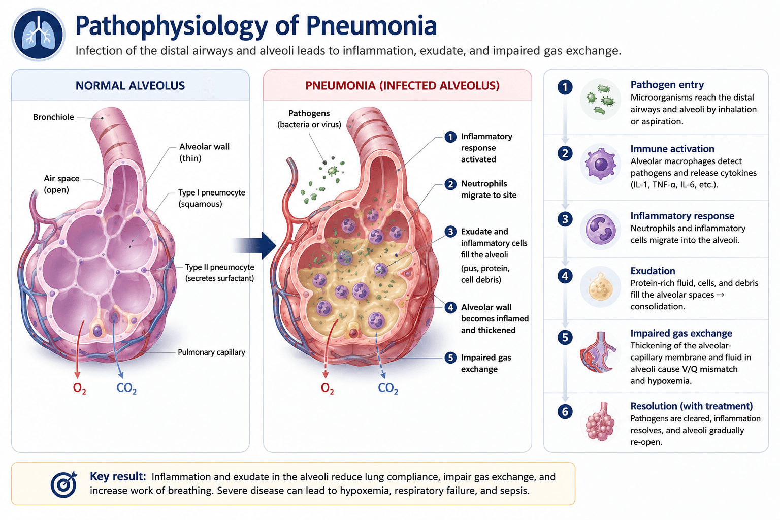
Pneumonia primarily involves the distal airways, alveoli, and pulmonary interstitium. In bacterial pneumonia, inflammatory material may fill alveolar spaces and produce consolidation. Viral infections more commonly create diffuse or patchy inflammation, although clinical and radiographic patterns frequently overlap.
Alveoli
Inflammatory cells, protein-rich fluid, and cellular debris may replace air within affected alveoli.
Interstitium
Inflammation may involve the supporting tissue surrounding alveoli and small airways.
Pleural space
Extension beyond the lung parenchyma may produce a parapneumonic effusion or empyema.
Pathophysiology
Pneumonia develops when a pathogen reaches the lower respiratory tract and overcomes normal host defenses, including mucociliary clearance, cough, airway immune responses, and alveolar macrophages.
Viral
- • Often affects younger children
- • May produce diffuse or patchy inflammation
- • Frequently accompanied by upper respiratory symptoms
- • Antibiotics do not treat the viral infection
Typical bacterial
- • Streptococcus pneumoniae is the most common bacterial cause
- • May present with abrupt fever and focal findings
- • Can produce alveolar consolidation
- • May progress to pleural complications
Atypical
- • More common in school-aged children and adolescents
- • Often associated with persistent cough
- • Mycoplasma pneumoniae and Chalmydophilia pneumoniae are common consideration
- • Clinical findings may be more diffuse
History and exam
Important history
- • Duration and progression of fever and cough
- • Breathing difficulty, chest pain, or exercise intolerance
- • Oral intake and urine output
- • Vomiting, lethargy, or altered mental status
- • Recent viral illness or sick contacts
- • Immunization status
- • Recent antibiotics or hospitalization
- • Aspiration risk or neurologic impairment
- • Tuberculosis exposure or travel history
- • Underlying cardiac, pulmonary, or immune disease
Examination priorities
- • General appearance and interaction
- • Respiratory rate measured while calm
- • Pulse oximetry
- • Retractions, nasal flaring, grunting, or head bobbing
- • Focal crackles or bronchial breath sounds
- • Decreased or asymmetric air entry
- • Signs of dehydration
- • Perfusion and mental status
Do not rely on auscultation alone
Children may have pneumonia without classic focal crackles. Respiratory rate, oxygen saturation, work of breathing, hydration, and overall appearance are often more important for determining severity.
| Pattern | Common clues | Important caution |
|---|---|---|
| Viral | Rhinorrhea, diffuse findings, wheezing, gradual onset | Viral and bacterial infection can coexist |
| Typical bacterial | Higher fever, focal crackles, focal decreased air entry | No single symptom confirms bacterial disease |
| Atypical | School age, persistent dry cough, headache, malaise | Presentation may overlap with viral disease |
Differential diagnosis
Viral bronchiolitis
Usually occurs in infants with diffuse crackles, wheezing, and upper respiratory symptoms.
Asthma exacerbation
Wheezing and prolonged expiration may predominate, often with a history of recurrent symptoms.
Foreign body aspiration
Consider with sudden onset, choking, unilateral wheezing, biphasic stridor, or asymmetric breath sounds.
Atelectasis
May produce focal opacity and reduced air entry, particularly with mucus plugging.
Pulmonary edema
Consider with cardiac disease, hepatomegaly, edema, or diffuse bilateral findings.
Tuberculosis
Consider with prolonged symptoms, weight loss, exposure history, or epidemiologic risk.
Pulmonary embolism
Uncommon in children but possible with thrombosis risk factors and pleuritic symptoms.
Malignancy or mass
Consider with recurrent focal pneumonia, persistent opacity, or systemic symptoms.
Aspiration
Consider with swallowing dysfunction, neurologic impairment, reflux, seizures, or altered consciousness.
Diagnostic workup
Pneumonia is often a clinical diagnosis. Testing should be guided by disease severity, diagnostic uncertainty, treatment failure, underlying conditions, and concern for complications.
Outpatient, uncomplicated disease
- • Measure vital signs and oxygen saturation
- • Chest radiograph is usually unnecessary
- • Routine CBC, CRP, and blood culture are unnecessary
- • Viral testing may be useful when it changes management
Hospitalized or complicated disease
- • Consider chest radiography
- • Consider CBC and inflammatory markers
- • Obtain cultures when clinically appropriate
- • Evaluate for sepsis or organ dysfunction
- • Use ultrasound when pleural fluid is suspected
| Test | When it may help | Limitation |
|---|---|---|
| Pulse oximetry | Assess severity and need for respiratory support | A normal value does not exclude pneumonia |
| Chest radiograph | Hypoxemia, significant distress, admission, failed treatment, or suspected complication | Cannot reliably determine viral versus bacterial etiology |
| Lung ultrasound | Evaluate suspected consolidation or pleural fluid | Operator dependent |
| CBC / CRP / procalcitonin | May support assessment of severe or complicated illness | Cannot independently distinguish bacterial from viral pneumonia |
| Blood culture | Severe disease, toxic appearance, complicated pneumonia, or selected hospitalized patients | Low yield in uncomplicated disease |
| Chest CT | Selected cases with unclear anatomy, necrosis, abscess, mass, or persistent disease | Radiation exposure; not routine |
Imaging pearl
Do not obtain a chest radiograph solely to prove pneumonia in a well-appearing child who can be safely treated as an outpatient. Imaging becomes more valuable when the diagnosis is uncertain, the child is significantly ill, or a complication is suspected.
Management
Management depends on suspected etiology, age, illness severity, immunization status, comorbidities, local resistance patterns, and ability to tolerate oral therapy.
Supportive care
- • Oxygen when hypoxemic
- • Oral or IV hydration as needed
- • Antipyretics for comfort
- • Nasal suctioning in younger children when helpful
- • Respiratory support based on work of breathing
- • Frequent reassessment for deterioration
Antibiotic principles
- • Avoid antibiotics when viral pneumonia is strongly favored
- • Use the narrowest effective agent
- • Transition from IV to oral therapy when improving
- • Adjust therapy to cultures and local antibiogram
- • Reassess if improvement does not occur as expected
| Clinical situation | Common approach | Key consideration |
|---|---|---|
| Suspected viral CAP | Supportive care | Antibiotics do not treat viral infection |
| Uncomplicated outpatient bacterial CAP | Oral amoxicillin is commonly first-line | Confirm allergy history, local pathway, and follow-up |
| Hospitalized uncomplicated bacterial CAP | IV ampicillin may be appropriate in a fully immunized child | Broaden based on severity, immunization status, or resistance risk |
| Severe or complicated CAP | Broader IV therapy with specialist and local-pathway guidance | Consider S. aureus coverage when clinically indicated |
| Suspected atypical pneumonia | Consider a macrolide in the appropriate age and clinical setting | Do not use cough alone to diagnose Mycoplasma |
Treatment duration
Many children with uncomplicated bacterial CAP who improve promptly can be treated with a short antibiotic course. Final agent, dose, and duration should follow the patient’s age, severity, response, local antibiogram, and institutional pathway.
Complications
Suspect complicated pneumonia when a child has persistent or worsening fever, escalating respiratory support, significant unilateral findings, chest pain, toxic appearance, or failure to improve after appropriate therapy.
Parapneumonic effusion
Fluid accumulates in the pleural space adjacent to pneumonia.
Clinical clues
- • Decreased breath sounds
- • Dullness to percussion
- • Persistent fever
- • Blunting or fluid on imaging
Empyema
Purulent or infected inflammatory material accumulates in the pleural space.
Clinical clues
- • Toxic appearance
- • Loculated pleural fluid
- • Persistent systemic inflammation
- • Failure to improve
Necrotizing pneumonia
Inflammation and tissue injury produce areas of pulmonary necrosis and cavitation.
Clinical clues
- • Severe or prolonged illness
- • Persistent fever
- • Cavitary changes
- • Complicated pleural disease
Imaging gallery
Imaging findings in complicated pneumonia
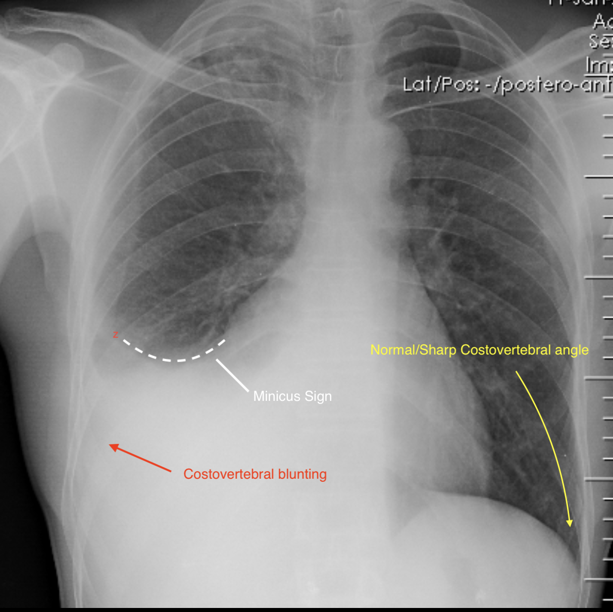
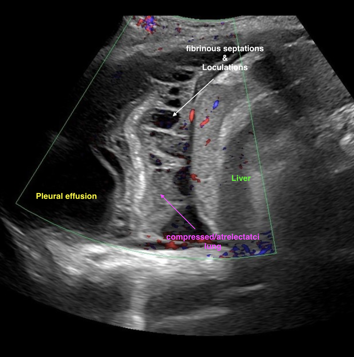
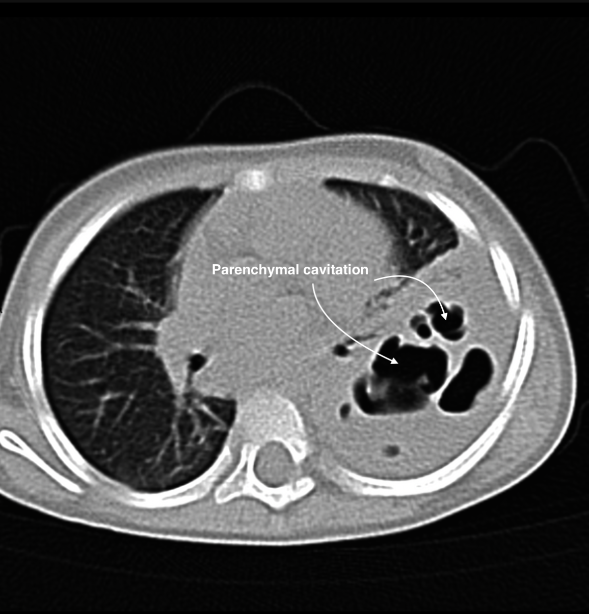
These examples demonstrate findings that may be seen with complicated pneumonia. Imaging findings are supportive rather than diagnostic in isolation because multiple pulmonary processes can produce similar appearances. Imaging should always be interpreted alongside the history, physical examination, laboratory findings, and clinical course.
Chest radiograph
Parapneumonic pleural effusion
Pleural fluid produces an upward-curving meniscus and blunting of the costophrenic angle adjacent to basilar airspace disease.
Radiopaedia Case: Pleural Effusion (Case 7)
Chest ultrasound
Complicated parapneumonic effusion / empyema
Internal echoes, septations, and loculations suggest an organized pleural infection and may influence drainage planning.
Radiopaedia Case: Thoracic Empyema (Case 8)
Chest CT
Necrotizing pneumonia
Multiple cavities embedded within consolidated lung suggest pulmonary necrosis and destruction of normal architecture.
Radiopaedia Case: Cavitating Pneumonia (Case 3)
Imaging is one piece of the clinical picture
Similar imaging findings may occur with infectious, inflammatory, congenital, and neoplastic processes. Imaging should be used to answer a focused clinical question and interpreted within the broader clinical context.
Evaluating pleural fluid
Chest ultrasound is particularly useful for confirming pleural fluid, estimating its size, and identifying septations or loculations. Management ranges from antibiotics and observation to drainage with a chest tube, fibrinolytic therapy, or surgery, depending on respiratory compromise and the character and size of the collection.
Disposition
Consider discharge
- • Well appearing
- • No significant hypoxemia
- • Minimal work of breathing
- • Tolerating oral fluids and medication
- • Reliable caregivers and follow-up
- • No concerning complication
Consider admission
- • Persistent oxygen requirement
- • Moderate or worsening respiratory distress
- • Inability to maintain hydration
- • Toxic appearance or concern for sepsis
- • Failed outpatient therapy
- • Complicated pneumonia
- • High-risk underlying condition
- • Unsafe or unreliable follow-up
Consider ICU care
- • Escalating oxygen requirement
- • Need for noninvasive or invasive ventilation
- • Apnea or exhaustion
- • Altered mental status
- • Hemodynamic instability
- • Severe sepsis or organ dysfunction
Return precautions
- • Increasing work of breathing
- • Cyanosis, apnea, or abnormal sleepiness
- • Inability to drink or markedly decreased urine output
- • Persistent vomiting
- • Worsening fever or clinical condition
- • Failure to begin improving within the expected timeframe
Clinical pearls
Interactive quiz
A fully immunized 5-year-old has fever, cough, tachypnea, focal right-sided crackles, and mild retractions. Oxygen saturation is 96% on room air. The child is drinking, alert, and has reliable follow-up. What is the most appropriate next step when bacterial community-acquired pneumonia is suspected?
Evidence Base
Clinical References
PIDS/IDSA pediatric community-acquired pneumonia guideline.
Evidence-based guideline for pediatric community-acquired pneumonia.
Pediatric Care Online clinical overview of pneumonia.
Global overview of pneumonia in children.
Educational Disclaimer
PediAtlas is intended for medical education and should not replace institutional guidelines, local protocols, patient-specific assessment, or clinical judgment.