Toxin-mediated respiratory infection
Pertussis
A highly contagious respiratory illness caused by Bordetella pertussis and characterized by prolonged cough, paroxysms, post-tussive vomiting, and potentially severe disease in young infants.
Clinical vignette
An infant with apnea and prolonged coughing spells
A 2-month-old infant presents with 10 days of worsening cough. The parents describe repeated coughing spells followed by facial color change, gasping, and occasional vomiting. The infant has had several brief pauses in breathing but little fever. Between episodes, the lung examination is relatively unremarkable.
Your first clinical task
Recognize that young infants may have severe pertussis without a classic whoop and may present primarily with apnea, cyanosis, or feeding difficulty.
Anatomy & localization
Pertussis targets the ciliated conducting airway
Bordetella pertussis attaches to ciliated respiratory epithelial cells, particularly within the trachea, bronchi, and larger bronchioles. As the airway becomes smaller, ciliated cells become progressively less abundant, while club cells assume a greater role in epithelial protection, secretion, detoxification, and repair.
Primary structures involved
- • Ciliated respiratory epithelium
- • Trachea
- • Main and segmental bronchi
- • Larger bronchioles
Secondary complications
- • Mucus plugging and atelectasis
- • Secondary bacterial pneumonia
- • Hypoxemia during severe paroxysms
- • Pulmonary hypertension in critical infant disease
Airway epithelial transition
Cilia become progressively less abundant down the bronchial tree
Trachea and bronchi
Pseudostratified ciliated columnar epithelium with mucus-producing goblet cells.
Abundant cilia move mucus, microorganisms, and trapped debris toward the pharynx through the mucociliary escalator.
Larger bronchioles
The lining transitions toward simple columnar or cuboidal epithelium, with fewer goblet cells and increasing numbers of club cells.
Ciliary clearance remains important, while club cells contribute protective secretions, detoxification, and epithelial repair.
Terminal bronchioles
Simple cuboidal epithelium containing club cells and relatively sparse ciliated cells.
This is the final portion of the conducting zone before the gas-exchanging airways begin.
Respiratory bronchioles
Low cuboidal epithelium with club cells and occasional proximal ciliated cells. Alveoli begin to interrupt the airway wall.
This region marks the transition from the conducting zone to the respiratory zone.
Alveolar ducts and alveoli
Predominantly thin type I pneumocytes, with type II pneumocytes producing surfactant and supporting epithelial repair.
The thin alveolar-capillary barrier permits efficient oxygen and carbon-dioxide exchange.
Why this matters in pertussis
Bordetella pertussis primarily attaches to ciliated respiratory epithelial cells. Damage to this mucociliary system impairs secretion clearance and contributes to retained mucus, airway irritation, and prolonged paroxysmal coughing.
Pathophysiology
Attachment, toxin production, and impaired clearance
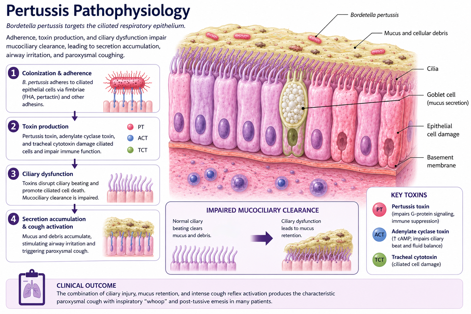
Pathophysiologic cascade
The four steps below mirror the infographic and summarize the causal chain from airway colonization to the characteristic clinical manifestations.
Attachment and colonization
B. pertussis adheres to ciliated respiratory epithelial cells through adhesins such as filamentous hemagglutinin, pertactin, and fimbriae, without deeply invading tissue.
Toxin production
Pertussis toxin, adenylate cyclase toxin, and tracheal cytotoxin impair immune defenses, disrupt epithelial function, and injure ciliated cells.
Ciliary dysfunction
Loss of coordinated ciliary beating impairs mucociliary clearance, allowing mucus and cellular debris to accumulate within the conducting airways.
Clinical consequences
Retained secretions and airway irritation trigger paroxysmal cough, inspiratory whoop, post-tussive emesis, cyanosis, and apnea in young infants.
Infant physiology matters
Young infants have limited respiratory reserve and may develop apnea, bradycardia, cyanosis, feeding intolerance, or respiratory failure without producing a classic inspiratory whoop.
Clinical stages
The illness evolves over several weeks
Catarrhal stage
Usually 1–2 weeks
Rhinorrhea, mild cough, lacrimation, and little or no fever. The illness is highly contagious and often resembles a routine viral upper respiratory infection.
Paroxysmal stage
Usually 2–6 weeks or longer
Repeated bursts of rapid coughing may be followed by an inspiratory whoop, cyanosis, exhaustion, or post-tussive vomiting. Young infants may present primarily with apnea.
Convalescent stage
Weeks to months
Coughing episodes gradually become less frequent and severe, but paroxysms may recur temporarily with subsequent respiratory infections (hence the term - 100-day cough).
History and physical exam
Listen for the pattern, not only the whoop
History
- • Prolonged or worsening cough
- • Repetitive paroxysms of coughing
- • Post-tussive vomiting
- • Whoop, gasping, cyanosis, or exhaustion
- • Apnea or bradycardia in young infants
- • Minimal or absent fever
- • Vaccination status and known exposure
- • Feeding tolerance and urine output
Physical exam
- • Respiratory rate and oxygen saturation
- • Work of breathing between and during episodes
- • Apnea, cyanosis, or altered responsiveness
- • Hydration and feeding ability
- • Focal findings suggesting pneumonia
- • Signs of exhaustion or respiratory failure
Differential diagnosis
A prolonged cough has many possible causes
Viral respiratory infection
Usually causes a shorter, less paroxysmal cough and may be accompanied by fever, congestion, or other viral symptoms.
Bronchiolitis
Typically affects infants with diffuse wheezing, crackles, increased work of breathing, and a viral prodrome.
Asthma
Recurrent wheezing, prolonged expiration, triggers, and improvement with bronchodilator therapy.
Pneumonia
Fever, focal crackles, focal decreased air entry, hypoxemia, or toxic appearance suggest pneumonia.
Foreign body aspiration
Abrupt choking or coughing onset with focal wheeze or asymmetric breath sounds should prompt consideration of aspiration.
Reflux or aspiration
Symptoms associated with feeds, choking, recurrent aspiration events, or chronic respiratory symptoms may suggest an alternative cause.
Diagnostic workup
Test early and collect the correct specimen
Nasopharyngeal PCR
PCR is commonly used because it provides rapid and sensitive detection. Yield is highest early and decreases as the cough progresses or after effective antibiotic therapy.
Culture
Culture is highly specific and useful for public-health surveillance, but sensitivity declines after the first weeks of illness and results take longer.
CBC
Leukocytosis with absolute lymphocytosis can support the diagnosis and may be pronounced in severe infant disease, but a normal CBC does not exclude pertussis.
Chest radiograph
Imaging is not routinely required in uncomplicated disease. Consider it when hypoxemia, focal findings, respiratory distress, or concern for pneumonia, atelectasis, or another diagnosis is present.
Do not delay treatment in a high-risk patient
Begin treatment before test confirmation when clinical suspicion is high and the patient is a young infant, pregnant, severely ill, or likely to expose someone at high risk for severe disease.
Management
Reduce transmission while supporting the child
Supportive care
- • Minimize unnecessary stimulation
- • Monitor oxygenation and apnea
- • Provide oxygen when hypoxemic
- • Support hydration and nutrition
- • Suction secretions when helpful
- • Escalate respiratory support when needed
Antimicrobial therapy
- • Macrolides are the first-line antibiotics for pertussis.
- • Azithromycin is generally the preferred agent, particularly in infants.
- • Treat early (during the catarrhal phase) to reduce symptom severity and transmission.
- • Follow age-specific recommendations and local guidance
Infection control and close contacts
- • Use appropriate droplet precautions
- • Notify infection prevention or public health as required
- • Assess household and other close contacts
- • Prioritize post-exposure prophylaxis for people at high risk and those likely to expose them
- • Review and update pertussis vaccination when indicated
Treatment expectation
Antibiotics are most effective when given early. Once the paroxysmal stage is established, the cough may continue for weeks despite successful eradication of the organism.
Disposition
Young infants deserve a low threshold for admission
Disposition depends on age, respiratory stability, feeding, hydration, comorbidities, and the caregiver's ability to recognize deterioration. Infants younger than 3 months warrant particularly cautious assessment, with the greatest risk occurring in those younger than 2 months.
Consider outpatient care
- • Infant at least 3 months old or older child with mild disease
- • No apnea, cyanosis, bradycardia, or significant hypoxemia
- • No meaningful respiratory distress between coughing episodes
- • Adequate feeding and hydration
- • No high-risk comorbidity or concerning laboratory findings
- • Reliable caregivers, return precautions, and close follow-up
Infants 3–6 months remain at increased risk. Age alone should not override concerning symptoms or an unreliable examination.
Strongly consider admission
- • Age younger than 3 months, especially younger than 2 months
- • Apnea, cyanosis, bradycardia, or post-tussive heart-rate decrease
- • Feeding difficulty, weight loss, or dehydration
- • Hypoxemia, pneumonia, or significant respiratory distress
- • Frequent, prolonged, or severe coughing paroxysms
- • Marked leukocytosis with lymphocytosis
- • Prematurity, cardiopulmonary disease, immunodeficiency, or concerning viral coinfection
- • Unsafe home observation or inability to ensure close follow-up
Escalate to intensive care
- • Recurrent or prolonged apnea
- • Respiratory failure or rapidly increasing respiratory support
- • Severe or recurrent hypoxemia
- • Hemodynamic instability
- • Pulmonary hypertension
- • Rapid clinical deterioration
- • Marked leukocytosis with critical infant disease
Clinical pearl
Young infants with pertussis may initially appear deceptively well, with only mild coryza or cough, before progressing to gagging, gasping, bradycardia, cyanosis, or apnea. A reassuring appearance early in the illness should not outweigh the patient's age, history, or observed events when deciding disposition.
Clinical pearls
High-yield pertussis takeaways
The whoop may be absent
Young infants may present with apnea, cyanosis, bradycardia, or feeding difficulty rather than a classic inspiratory whoop.
Minimal fever is a clue
Prominent fever should prompt consideration of pneumonia, another infection, or a complication.
Post-tussive vomiting is highly suggestive
Vomiting immediately after repetitive coughing spells is a classic clinical clue.
Vaccination modifies disease
Vaccinated children and adults may still develop pertussis but often have less classic or less severe symptoms.
Late antibiotics do not immediately stop the cough
Once toxin-mediated airway dysfunction and paroxysms are established, symptoms may persist even after bacterial eradication.
Protect vulnerable contacts
Management includes the patient, infection control, contact assessment, prophylaxis decisions, and vaccination review.
Knowledge check
Which finding most strongly suggests pertussis?
A 6-month-old has had a worsening cough for 12 days with little fever. Which additional finding would most strongly support the diagnosis of pertussis?
Evidence Base
Clinical References
Clinical care guidance for treatment, postexposure prophylaxis, and management of pertussis.
Red Book guidance for the diagnosis, treatment, prevention, and infection control of pertussis.
Clinical review of pertussis presentation, pathophysiology, diagnosis, and management in children.
Primary literature regarding severe pertussis in young infants, including apnea, leukocytosis, and cardiopulmonary complications.
Secondary Educational Sources
UpToDate and OpenEvidence were used for educational verification and consistency checks where appropriate.
Educational Disclaimer
PediAtlas is intended for medical education and should not replace institutional guidelines, local protocols, patient-specific assessment, or clinical judgment.