Mechanical airway obstruction
Foreign Body Aspiration
Entry of food or another object into the larynx, trachea, or bronchial tree can cause complete obstruction, focal air trapping, atelectasis, or delayed recurrent pulmonary disease. A child may appear well after the initial choking event despite a retained airway foreign body.
Clinical vignette
A toddler with sudden choking and unilateral wheezing
A previously healthy 22-month-old boy is brought to the emergency department after suddenly coughing and choking while eating peanuts. His parents report that he briefly became cyanotic but quickly recovered. Since the event, he has continued to cough intermittently. Examination reveals mild tachypnea, decreased breath sounds, and an expiratory wheeze over the right lung field. Oxygen saturation is 97% on room air.
Your first clinical task
Determine whether the object entered the airway or the esophagus, assess for current airway compromise, and recognize that normal oxygenation does not exclude a retained bronchial foreign body.
Anatomy & localization
Airway or esophagus?
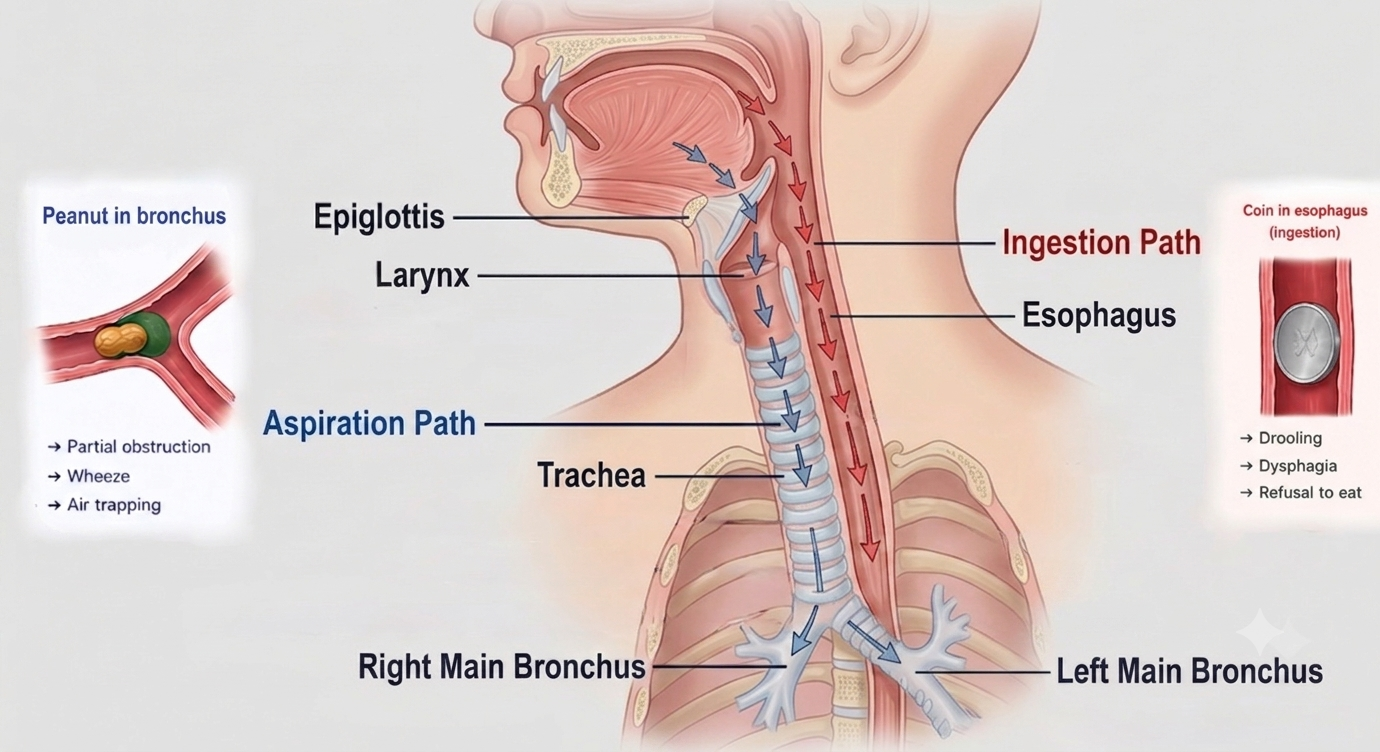
After passing through the oropharynx, an object may enter the anterior laryngeal inlet and tracheobronchial tree or pass posteriorly into the esophagus. Symptoms depend on both the pathway taken and the level at which the object becomes lodged.
Airway aspiration
Think cough, wheeze, or stridor
- • Sudden choking or coughing
- • Inability to speak or cry with complete obstruction
- • Stridor or voice change with upper-airway impaction
- • Unilateral wheeze or decreased breath sounds
- • Persistent cough after the child appears to recover
- • Recurrent focal pneumonia with delayed diagnosis
Esophageal ingestion
Think drooling, dysphagia, or feeding refusal
- • Drooling or inability to manage secretions
- • Dysphagia or odynophagia
- • Gagging, vomiting, or feeding refusal
- • Retrosternal or neck discomfort
- • Object visible within the esophagus on imaging
- • Possible respiratory symptoms from posterior tracheal compression
Localization within the airway
Larynx or subglottis
Stridor, hoarseness, aphonia, severe distress, or complete airway obstruction.
Trachea
Cough, biphasic noisy breathing, variable wheeze, or an object that may move with changes in position.
Mainstem bronchus
Unilateral wheeze, asymmetric breath sounds, focal air trapping, or atelectasis.
Distal bronchus
Subtle focal findings, persistent cough, segmental atelectasis, or recurrent pneumonia in the same location.
Pediatric anatomy pearl
Do not assume every pediatric foreign body enters the right mainstem bronchus. Unlike adults, young children have less pronounced differences between the right and left mainstem bronchial angles, so either side may be involved.
Pathophysiology
The object changes airflow and obstructs secretion clearance
The physiologic consequences depend on the object's size, shape, location, duration, and whether air can pass around it during inspiration or expiration.
Complete obstruction
The object completely blocks airflow. A child may be unable to speak, cough effectively, or breathe and can rapidly develop cyanosis, loss of consciousness, and cardiac arrest.
Partial fixed obstruction
Airflow is reduced during both inspiration and expiration. Depending on location, this may produce stridor, persistent wheeze, decreased air entry, or respiratory distress.
Ball-valve obstruction
Air enters around the object during inspiration but cannot escape effectively during expiration, producing distal air trapping and unilateral hyperinflation.
Delayed distal disease
A retained foreign body causes mucosal edema, impaired secretion clearance, granulation tissue, atelectasis, recurrent pneumonia, or bronchiectasis.
Deceptive improvement
The dramatic choking episode may stop once the object moves distally. This apparent recovery does not confirm expulsion—the child may enter a quieter interval despite persistent bronchial obstruction.
History and physical exam
Ask specifically about the moment symptoms began
History
- • Sudden coughing, choking, gagging, or cyanosis
- • Eating or playing with small objects at onset
- • Witnessed versus unwitnessed event
- • Type, size, shape, and number of possible objects
- • Persistent cough, wheeze, stridor, or voice change
- • Drooling, dysphagia, vomiting, or feeding refusal
- • Fever or recurrent focal pneumonia
- • Response—or lack of response—to asthma therapy
Physical exam
- • Ability to speak, cry, and cough effectively
- • Mental status, color, and oxygen saturation
- • Stridor or altered voice
- • Respiratory rate and work of breathing
- • Unilateral wheeze or asymmetric air entry
- • Focal crackles or signs of atelectasis
- • Drooling and ability to manage secretions
- • Signs of exhaustion or impending respiratory failure
The examination may be normal
Children with a retained foreign body may appear comfortable between episodes and may have normal oxygen saturation, no audible wheeze, or subtle findings. Clinical suspicion must incorporate the history rather than relying on one reassuring examination.
Differential diagnosis
Sudden focal symptoms are the strongest clue
Esophageal foreign body
Drooling, dysphagia, odynophagia, feeding refusal, vomiting, or retrosternal discomfort suggest ingestion. A large esophageal object can still cause cough, stridor, or respiratory symptoms through posterior tracheal compression.
Asthma
Asthma usually causes recurrent or diffuse wheezing and improves with bronchodilator therapy. Sudden onset, a witnessed choking event, or persistent focal wheeze argues against uncomplicated asthma.
Bronchiolitis
A viral prodrome followed by diffuse bilateral wheezing or crackles is more typical. Abrupt onset and asymmetric breath sounds should raise concern for an aspirated object.
Pneumonia
Fever, focal crackles, hypoxemia, and an acute infectious course support pneumonia. Recurrent pneumonia in the same lobe may be the delayed presentation of a retained foreign body.
Croup or upper-airway infection
Barking cough, hoarseness, and viral symptoms suggest croup. Abrupt stridor while eating or playing with a small object should prompt concern for a laryngeal or tracheal foreign body.
Anaphylaxis
Rapid respiratory symptoms accompanied by urticaria, angioedema, vomiting, hypotension, or multisystem involvement suggest anaphylaxis rather than mechanical obstruction.
Diagnostic workup
Normal imaging does not exclude aspiration
Standard radiographs
Obtain 2-view chest radiographs when the child is stable. Imaging may directly show a radiopaque object or indirectly demonstrate unilateral hyperinflation, mediastinal shift, atelectasis, consolidation, or pneumothorax.
Additional imaging in selected patients
Standard frontal and lateral chest radiographs remain the initial imaging study for suspected foreign body aspiration. Inspiratory-expiratory or lateral decubitus views have historically been used to demonstrate unilateral air trapping, but their diagnostic performance is limited and they are no longer routinely recommended in many centers.
Imaging for possible ingestion
When the pathway or object is uncertain, imaging may need to include the neck, chest, and abdomen. Coins, button batteries, magnets, sharp objects, and other high-risk ingestions require object-specific urgent management.
CT in selected patients
Most aspirated foreign bodies are radiolucent, and normal chest radiographs do not exclude aspiration. When suspicion remains high despite unrevealing radiographs, further evaluation should not be delayed. Depending on institutional practice and resource availability, patients may proceed directly to bronchoscopy or undergo low-dose noncontrast chest CT when the diagnosis remains uncertain.
Imaging Pearls
Review these classic imaging findings. Study the image first, then reveal the annotations to identify the important features.
Airway Foreign Body
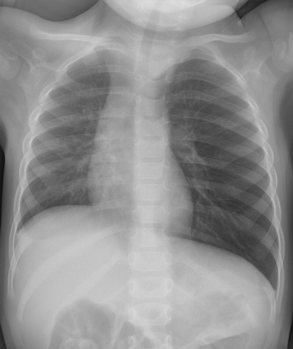
Wikimedia Commons: Foreign Body Aspiration Chest Radiograph (Hellerhoff, 2022) · CC BY-SA 4.0
Clinical Pearl
Most aspirated foreign bodies are radiolucent and are not directly visible on chest radiographs. Diagnosis often relies on indirect findings such as unilateral hyperinflation, air trapping, and mediastinal shift.
Esophageal Coin
AP and lateral radiographs demonstrate the typical appearance of an esophageal foreign body. The lateral view confirms that the coin lies posterior to the trachea.
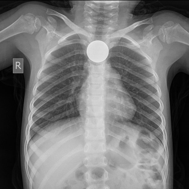
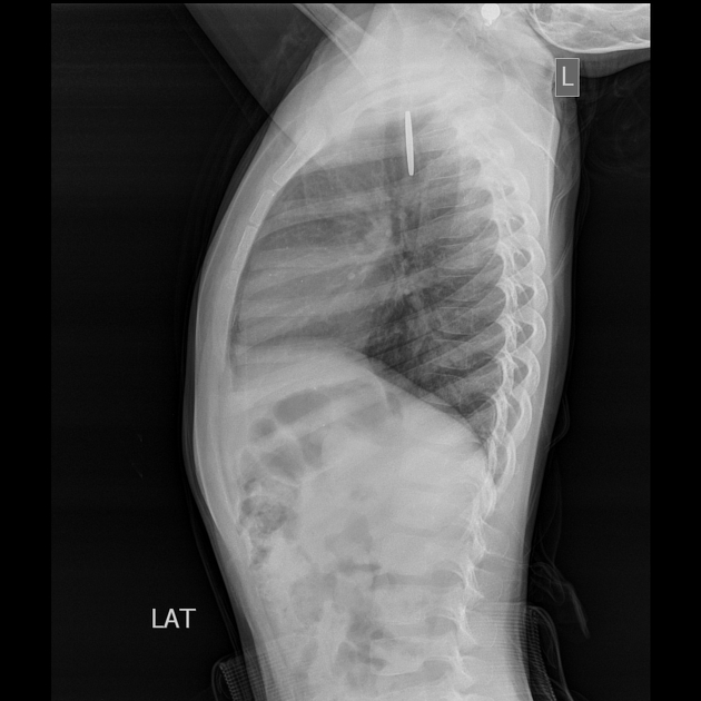
🔍 Click either image to view the full-resolution radiograph.
Radiopaedia Case: Oesophageal Foreign Body – Coin. Licensed under CC BY-NC-SA.
Clinical Pearl
- On the AP radiograph, esophageal coins are usually seen en face (round), whereas tracheal coins more commonly appear edge-on.
- The lateral radiograph confirms the coin is posterior to the trachea, supporting an esophageal location.
- Always distinguish a coin from a button battery, which requires emergent removal because of the risk of caustic injury.
Bronchoscopy is the definitive diagnostic test
If clinical suspicion remains high, urgent consultation for bronchoscopy is appropriate even when radiographs are normal. Bronchoscopy allows direct airway visualization and removal during the same procedure.
Management
Management depends on whether obstruction is complete
Severe active airway obstruction
- • Activate emergency assistance immediately
- • Allow coughing when the cough remains strong and effective
- • For an infant: cycles of 5 back blows and 5 chest thrusts (heel-of-hand technique)
- • For a child: cycles of 5 back blows and 5 abdominal thrusts
- • If unresponsive, begin CPR starting with compressions
- • Remove an object only when it is clearly visible
- • Do not perform a blind finger sweep
Stable suspected retained foreign body
- • Keep the child calm and minimize agitation
- • Provide oxygen when indicated
- • Maintain continuous clinical observation
- • Keep the child NPO
- • Avoid unnecessary airway manipulation
- • Obtain appropriate imaging if it will not delay care
- • Consult the local airway team urgently for bronchoscopy
Definitive removal
Rigid bronchoscopy under general anesthesia is commonly used because it provides airway control, ventilation, direct visualization, and access for extraction instruments. The exact procedural team varies by institution and may include otolaryngology, pediatric surgery, pulmonology, and anesthesia.
Avoid preventable deterioration
A partially obstructing object can move and suddenly produce complete obstruction. Do not leave the child unobserved or create unnecessary distress while awaiting definitive evaluation.
Disposition
Suspected airway foreign bodies require definitive planning
Consider discharge
- • Aspiration has been confidently excluded or the object was completely removed
- • Stable respiratory status and oxygenation
- • No stridor, focal wheeze, or asymmetric air entry
- • Able to tolerate appropriate oral intake
- • No procedural complication requiring observation
- • Clear return precautions and follow-up plan
Observe or admit
- • Ongoing concern for retained foreign body
- • Procedure planned but not immediately performed
- • Persistent cough, wheeze, or asymmetric examination
- • Oxygen requirement or increased work of breathing
- • Atelectasis, pneumonia, or other pulmonary complication
- • Post-bronchoscopy monitoring is indicated
Escalate immediately
- • Complete or rapidly worsening airway obstruction
- • Altered mental status or exhaustion
- • Severe hypoxemia or cyanosis
- • Marked stridor or minimal air movement
- • Increasing respiratory support
- • Hemodynamic instability or cardiac arrest
After removal
Reassess respiratory symptoms and examination findings. Depending on the duration and complexity of impaction, evaluate for residual fragments, airway edema, bleeding, granulation tissue, pneumothorax, atelectasis, or secondary infection.
Clinical pearls
High-yield foreign body aspiration takeaways
The choking event may be remote
Ask specifically about sudden coughing or choking even when symptoms began days or weeks earlier.
A normal radiograph does not rule it out
Most food items and many small objects are radiolucent, and both the examination and imaging may initially appear normal.
Unilateral wheeze is not always asthma
Sudden focal wheezing or persistent asymmetry—especially without prior recurrent symptoms—should prompt evaluation for aspiration.
Normal oxygen saturation can be misleading
A child may oxygenate normally despite significant focal bronchial obstruction.
Either mainstem bronchus may be involved
The right-sided predominance seen in adults is less reliable in young children.
Bronchoscopy can diagnose and treat
When suspicion remains high, direct visualization permits confirmation and removal during the same procedure.
Do not forget the esophagus
Drooling, dysphagia, feeding refusal, and vomiting suggest ingestion, but esophageal objects may also produce respiratory symptoms.
Knowledge check
What is the next best step?
A 2-year-old suddenly choked while eating nuts. He now appears comfortable with an oxygen saturation of 98%, but has persistent unilateral wheezing. Chest radiographs show no visible foreign body and no definite abnormality. What is the most appropriate next step?
Evidence Base
Clinical References
Current Pediatrics in Review discussion of the presentation, evaluation, and management of ingested and aspirated foreign bodies in children.
2025 pediatric basic life support guidance, including management of severe foreign-body airway obstruction in infants and children.
Clinical guidance for recognition, investigation, observation, consultation, and management of inhaled foreign bodies.
Overview of pediatric foreign body aspiration and bronchoscopic removal using a rigid ventilating bronchoscope.
Secondary Educational Sources
Additional AAP educational material and current institutional pediatric airway guidance were used for educational verification and consistency checks.
Educational Disclaimer
PediAtlas is intended for medical education and should not replace institutional guidelines, local protocols, patient-specific assessment, or clinical judgment.