Lower Airway Disease
Bronchiolitis
Acute viral inflammation of the bronchioles causing airway edema, mucus production, air trapping, and variable small-airway obstruction.
Clinical vignette
An infant with cough, congestion, and increased work of breathing
A previously healthy 7-month-old presents on day four of illness with rhinorrhea, cough, decreased feeding, tachypnea, subcostal retractions, diffuse crackles, and expiratory wheezing. Oxygen saturation is 91% on room air. The infant is alert but has taken only half of the usual feeding volume.
What should you decide first?
Determine respiratory severity, hydration status, risk factors for deterioration, and whether supportive care can be provided safely at home.
Anatomy localization
The bronchioles
The disease localizes to the small, non-cartilaginous airways. Their narrow baseline diameter makes infants particularly vulnerable to obstruction from edema, mucus, and cellular debris.
Edema
Thickens the bronchiolar wall.
Mucus
Narrows and plugs the airway lumen.
Air trapping
Occurs when expiration is more limited than inspiration.
Pathophysiology
From viral infection to respiratory distress
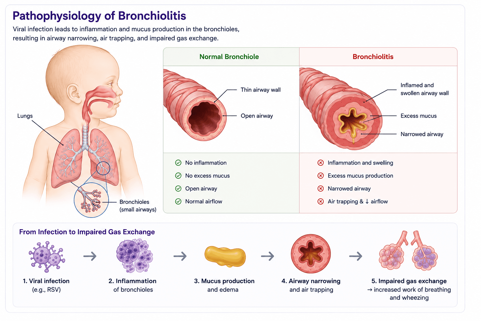
Viral infection of the respiratory epithelium
Inflammation, epithelial injury, and bronchiolar edema
Increased mucus and cellular debris
Partial or complete small-airway obstruction
Air trapping, atelectasis, ventilation-perfusion mismatch, and increased work of breathing
History and physical exam
Assess severity, not just the diagnosis
History
- • Day of illness and trajectory
- • Feeding volume and urine output
- • Apnea, cyanosis, or fatigue
- • Prematurity or cardiopulmonary disease
- • Age, especially young infants
- • Prior wheezing or strong atopic history
Physical exam
- • Respiratory rate and work of breathing
- • Retractions, nasal flaring, or grunting
- • Crackles, wheezing, and air entry
- • Oxygen saturation and mental status
- • Hydration and perfusion
- • Focal findings suggesting an alternative diagnosis
Differential diagnosis
What else could look like this?
Asthma or viral-induced wheeze
Recurrent episodes, prior response to bronchodilator, atopy, or older age.
Pneumonia
Focal examination findings, persistent high fever, or toxic appearance.
Foreign body aspiration
Abrupt onset, asymmetric air entry, focal wheeze, or choking history.
Heart failure
Hepatomegaly, diaphoresis with feeds, poor growth, murmur, or cardiomegaly.
Pertussis
Paroxysmal cough, apnea, post-tussive emesis, or minimal fever.
Diagnostic workup
Typical bronchiolitis requires very little testing
Usually appropriate
- • Focused history
- • Repeated respiratory assessment
- • Hydration assessment
- • Pulse oximetry when clinically indicated
Avoid routinely
- • Chest radiograph
- • Respiratory viral panel
- • CBC or inflammatory markers
- • Albuterol
- • Nebulized epinephrine
- • Systemic corticosteroids
- • Antibiotics without suspected bacterial infection
- • Chest physiotherapy
Testing may be appropriate when the presentation is atypical, severe, prolonged, focal, or when another diagnosis would change management.
Management
Supportive care first
Nasal suctioning
Use gentle, targeted suctioning when secretions impair breathing or feeding.
Hydration
Continue oral feeds when safe; consider enteral or intravenous support when intake is inadequate.
Oxygen
Provide supplemental oxygen for persistent clinically significant hypoxemia according to local policy.
Respiratory support
Escalate support when work of breathing, oxygenation, apnea, or fatigue worsens.
Oxygen threshold note
Many bronchiolitis guidelines use a persistent oxygen saturation below approximately 90% as a threshold for supplemental oxygen, but institutional policies and patient comorbidities may differ.
Disposition
Admit the child, not the pulse oximeter
Consider admission
- • Persistent hypoxemia
- • Moderate or severe work of breathing
- • Apnea, exhaustion, or altered responsiveness
- • Inadequate feeding or dehydration
- • High-risk age or underlying disease
- • Unsafe home monitoring or limited follow-up
Reasonable discharge
- • Stable or improving respiratory effort
- • Adequate oxygenation on room air
- • Sufficient oral intake
- • Caregiver can perform basic nasal care
- • Clear return precautions
- • Appropriate follow-up access
Clinical pearls
High-yield takeaways
Bronchiolitis is primarily a clinical diagnosis.
Supportive care is the foundation of management.
Suctioning is most useful when nasal obstruction interferes with feeding or breathing.
Routine chest radiography and viral testing are usually unnecessary.
Routine albuterol, systemic corticosteroids, and antibiotics are not recommended.
Knowledge check
What is the best next step?
A comfortable 8-month-old with typical bronchiolitis has mild retractions, adequate feeding, diffuse wheezing and crackles, and an oxygen saturation of 95% on room air. Which intervention is most appropriate?
Evidence Base
Clinical References
Diagnosis, management, and prevention of bronchiolitis.
Bronchiolitis: Recommendations for diagnosis, monitoring, and management.
Secondary Educational Sources
UpToDate and OpenEvidence were used for educational verification and consistency checks where appropriate.
Educational Disclaimer
PediAtlas is intended for medical education and should not replace institutional guidelines, local protocols, patient-specific assessment, or clinical judgment.