Chronic Lower Airway Disease
Asthma
A heterogeneous chronic respiratory disease characterized by variable symptoms and variable expiratory airflow limitation.
Clinical vignette
A child with recurrent cough and wheezing
An 8-year-old presents with cough, chest tightness, and wheezing after several days of rhinorrhea. He has nighttime cough twice weekly and develops similar symptoms with exercise. Examination reveals tachypnea, subcostal retractions, diffuse expiratory wheezing, and prolonged expiration.
Your first clinical task
Assess the severity of the current exacerbation while also recognizing the pattern of chronic asthma symptoms.
Anatomy localization
Asthma is a disease of the conducting airways
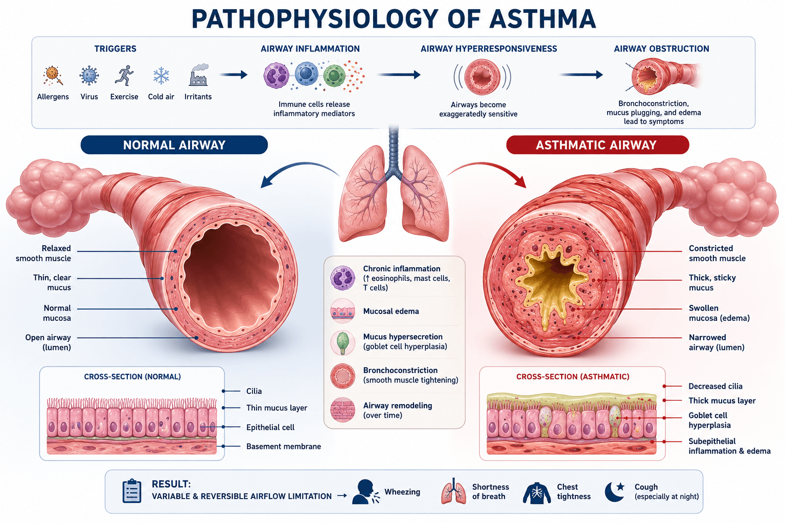
Asthma primarily affects the medium and small bronchi through inflammation, bronchial smooth muscle constriction, and mucus production resulting in variable airflow obstruction.
Structures involved
- • Medium and small bronchi
- • Bronchial smooth muscle
- • Airway mucosa
- • Goblet cells and mucus glands
Structures usually spared
- • Alveoli
- • Pleural space
- • Pulmonary vasculature
- • Lung parenchyma
Clinical implication
Because asthma affects the conducting airways rather than the alveoli, patients typically present with wheezing, prolonged expiration, chest tightness, and airflow obstruction rather than focal crackles or consolidation.
Pathophysiology
Inflammation plus reversible airway narrowing
Airway inflammation
Chronic inflammation makes the airways hyperresponsive to triggers.
Bronchoconstriction
Airway smooth muscle contracts, producing variable airflow obstruction.
Mucus production
Increased secretions further narrow the airway lumen.
Air trapping
Expiration becomes prolonged and incomplete, causing hyperinflation.
History and physical exam
Confirm the pattern and identify risk
History
- • Episodic wheeze, cough, dyspnea, or chest tightness
- • Nighttime or early-morning symptoms
- • Exercise, viral, allergen, smoke, or weather triggers
- • Prior emergency visits, hospitalization, or intubation
- • Controller adherence and inhaler technique
- • Atopy, eczema, allergic rhinitis, and family history
Physical exam
- • Respiratory rate and oxygen saturation
- • Retractions and accessory muscle use
- • Ability to speak, feed, or ambulate
- • Wheezing and prolonged expiration
- • Air entry, including a concerning silent chest
- • Mental status and signs of fatigue
Acute severity
How sick is the child right now?
Mild
Speaks normally, mild wheeze or retractions, good air movement.
Moderate
Increased work of breathing, more persistent wheeze, reduced activity or speech.
Severe
Marked retractions, difficulty speaking, poor air entry, agitation, or hypoxemia.
Impending respiratory failure
Fatigue, altered mental status, cyanosis, silent chest, or worsening despite treatment.
Future feature: an interactive severity tool using work of breathing, speech, oxygenation, air entry, and mental status.
Differential diagnosis
Not every wheezing child has asthma
Bronchiolitis
Usually a first episode in an infant with viral symptoms, crackles, and diffuse wheezing.
Foreign body aspiration
Abrupt onset, choking history, asymmetric air entry, or focal wheeze.
Pneumonia
Fever, focal crackles, focal decreased air entry, or toxic appearance.
Anaphylaxis
Acute respiratory symptoms with urticaria, angioedema, hypotension, or gastrointestinal symptoms.
Vocal cord dysfunction
Inspiratory symptoms, throat tightness, stridor, or poor response to asthma therapy.
Diagnostic workup
Confirm variable airflow obstruction and avoid unnecessary testing
Asthma is diagnosed by a characteristic pattern of variable respiratory symptoms together with objective evidence of variable expiratory airflow limitation when testing is feasible.
Initial evaluation
- • Detailed symptom and trigger history
- • Prior exacerbations and response to treatment
- • Physical examination and oxygen saturation
- • Assessment of atopy and relevant comorbidities
- • Review of inhaler technique and medication adherence
Objective lung-function testing
- • Perform spirometry with bronchodilator testing when the child is developmentally able
- • Look for reduced expiratory airflow before treatment
- • Repeat testing after an inhaled bronchodilator
- • Improvement in airflow supports variable, reversible obstruction
- • Normal spirometry between episodes does not completely exclude asthma
When the diagnosis remains uncertain
Additional options
- • Repeat spirometry during symptoms
- • Serial peak expiratory flow measurements
- • Exercise or bronchoprovocation testing
- • Specialist-directed allergy evaluation
Fractional exhaled nitric oxide
FeNO may support the diagnosis of type 2 airway inflammation when asthma remains uncertain, but it should be interpreted as an adjunct rather than used alone to diagnose or exclude asthma.
Avoid routinely in a typical exacerbation
- • Chest radiograph
- • CBC or inflammatory markers
- • Respiratory viral testing
- • Blood cultures
- • Arterial or venous blood gas
- • CT imaging
Consider targeted testing when
- • Findings are focal or markedly asymmetric
- • Foreign body aspiration is possible
- • Pneumonia or pneumothorax is suspected
- • The diagnosis is uncertain or the course is atypical
- • The child fails to improve as expected
- • Respiratory failure or another complication is suspected
Severe-asthma warning
A normal or rising carbon dioxide level may be ominous
Early in an asthma exacerbation, children commonly hyperventilate and lower their carbon dioxide level. In a child with severe distress, a normalizing or rising carbon dioxide level may indicate worsening fatigue and inadequate ventilation.
Decreasing wheeze with worsening air movement may represent a silent chest—not clinical improvement.
Management overview
Treat bronchoconstriction and inflammation
Short-acting bronchodilator
Administer inhaled albuterol for acute bronchoconstriction.
Systemic corticosteroid
Use early for moderate or severe exacerbations or incomplete response to initial bronchodilator therapy.
Oxygen
Provide supplemental oxygen when hypoxemia is present.
Escalation
Consider repeated or continuous bronchodilator therapy, ipratropium, magnesium, and higher-level respiratory support according to severity and local pathway.
Chronic-care principle
Asthma management should include an inhaled corticosteroid-containing strategy, assessment of symptom control and future risk, inhaler technique, adherence, trigger reduction, and a written asthma action plan.
Disposition
Reassess the response—not just the initial appearance
Disposition depends on the child's clinical response after initial treatment, oxygen requirement, work of breathing, air movement, ability to speak or feed, prior high-risk history, and the safety of outpatient follow-up.
Consider discharge
- • Symptoms and work of breathing are substantially improved
- • Air movement is good without concerning fatigue
- • Oxygenation is stable on room air
- • Bronchodilator treatments can be spaced appropriately
- • The child can speak, drink, and ambulate near baseline
- • Caregivers understand treatment and return precautions
- • Reliable follow-up and access to medications are available
Consider hospital admission
- • Persistent moderate or severe respiratory distress
- • Continued oxygen requirement or recurrent hypoxemia
- • Need for frequent or continuous bronchodilator therapy
- • Incomplete or short-lived response to initial treatment
- • Poor oral intake, dehydration, or inability to manage at home
- • High-risk history or significant comorbidity
- • Unsafe social situation or unreliable follow-up
Escalate to intensive care
- • Worsening despite aggressive initial therapy
- • Markedly reduced air movement or silent chest
- • Exhaustion, confusion, drowsiness, or agitation
- • Persistent or worsening hypoxemia
- • Rising carbon dioxide or evidence of ventilatory failure
- • Need for advanced or rapidly escalating respiratory support
- • Concern for impending respiratory arrest
Discharge bundle
Stabilization is only half of the job
Confirm an inhaled corticosteroid-containing treatment plan
Provide an appropriate reliever plan
Complete the prescribed systemic corticosteroid course when indicated
Demonstrate inhaler and spacer technique
Review triggers, adherence, and barriers to obtaining medication
Provide a written asthma action plan
Give clear return precautions
Arrange timely outpatient follow-up
High-risk history
Prior intensive-care admission, previous intubation, recent emergency visits or systemic corticosteroid courses, poor controller adherence, medication-access barriers, and psychosocial concerns should lower the threshold for observation, admission, or specialist involvement.
Exact admission, oxygen, and bronchodilator-spacing criteria vary by institution. Apply the local pediatric asthma pathway when making clinical decisions.
Clinical pearls
High-yield asthma takeaways
Wheezing intensity does not equal disease severity
A child with minimal wheezing but very poor air movement may be more critically ill than a child with loud wheezing and good air exchange.
A silent chest is an emergency
Diminishing wheeze accompanied by worsening work of breathing, fatigue, altered mental status, or poor air movement suggests critically limited airflow—not improvement.
Treat before ordering routine tests
A typical asthma exacerbation is assessed clinically, and urgent bronchodilator treatment should not be delayed for routine chest radiography or laboratory testing.
Look beyond the current exacerbation
Ask about nighttime symptoms, exercise limitation, prior emergency visits, systemic steroid courses, hospitalization, intensive-care admission, and previous intubation.
Inhaler technique is part of the treatment
Before escalating long-term therapy, confirm that the medication is being taken, the inhaler technique is correct, and the child has an appropriate spacer.
Asthma control and asthma severity are not the same
Control describes the child's current symptoms and future risk, while severity is judged retrospectively by the treatment required to maintain control.
Every child needs a plan after stabilization
Discharge planning should address controller therapy, reliever use, trigger reduction, spacer technique, follow-up, and a written asthma action plan.
Do not miss
Less wheezing can mean less airflow.
Always interpret wheezing together with air entry, respiratory effort, speech, oxygenation, fatigue, and mental status.
Knowledge check
What is the best next step?
A 9-year-old with known asthma presents with diffuse wheezing, moderate intercostal retractions, and difficulty speaking in full sentences. Oxygen saturation is 91% on room air. Which initial treatment is most appropriate?
Evidence Base
Clinical References
Global Strategy for Asthma Management and Prevention, 2026.
2020 Focused Updates to the Asthma Management Guidelines.
Educational Disclaimer
PediAtlas is intended for medical education and should not replace institutional guidelines, local protocols, patient-specific assessment, or clinical judgment.