Respiratory Foundations · Module 01
Anatomy & Localization
Follow air from the upper airway to the alveoli, map the lungs and pleural spaces, and connect respiratory findings to the anatomy producing them.
Core idea
Before you can understand respiratory disease, you need to know where the problem lives.
Start with the pathway
The Respiratory Map
Air travels through a branching system that transitions from conducting airways to specialized gas-exchanging units.
Mental Model
Follow one breath
Instead of memorizing respiratory structures as an isolated list, follow a breath from the nose and mouth through the conducting airways until it reaches the alveoli.
Airflow pathway
Nose & oral cavity
Entry points for inspired air. The nasal passages also warm, humidify, and filter incoming air.
Pharynx & larynx
The upper airway channels air toward the trachea while the larynx protects the entrance to the lower respiratory tract.
Trachea
The central conducting airway connecting the larynx to the right and left main bronchi.
Bronchi
The main bronchi enter the lungs and repeatedly branch into progressively smaller conducting airways.
Bronchioles
Small airways that lack the cartilaginous support of the larger bronchi and ultimately lead to the terminal bronchioles.
Respiratory bronchioles & alveolar ducts
The transition into the respiratory zone, where alveoli begin appearing along the airway walls.
Alveoli
Thin-walled gas-exchanging units closely associated with the pulmonary capillary network.
Conducting Zone
The conducting airways move air toward and away from the gas-exchanging portion of the lung. They include the larger airways and extend distally to the terminal bronchioles.
Primary job: move and condition air.
Respiratory Zone
Distal airways transition into respiratory bronchioles, alveolar ducts, and alveoli, where inspired gas comes into close contact with the pulmonary capillary circulation.
Primary job: gas exchange.
Localization Pearl
The level of obstruction changes the clinical picture
A problem at the larynx does not sound like a problem in the bronchioles. Learning the airway as a continuous pathway gives you the framework for later localizing findings such as stridor, wheezing, and focal loss of airflow.
Gross anatomy
Lungs, Lobes & Fissures
The lungs are divided into lobes by fissures, creating clinically useful regions for describing where disease is occurring.
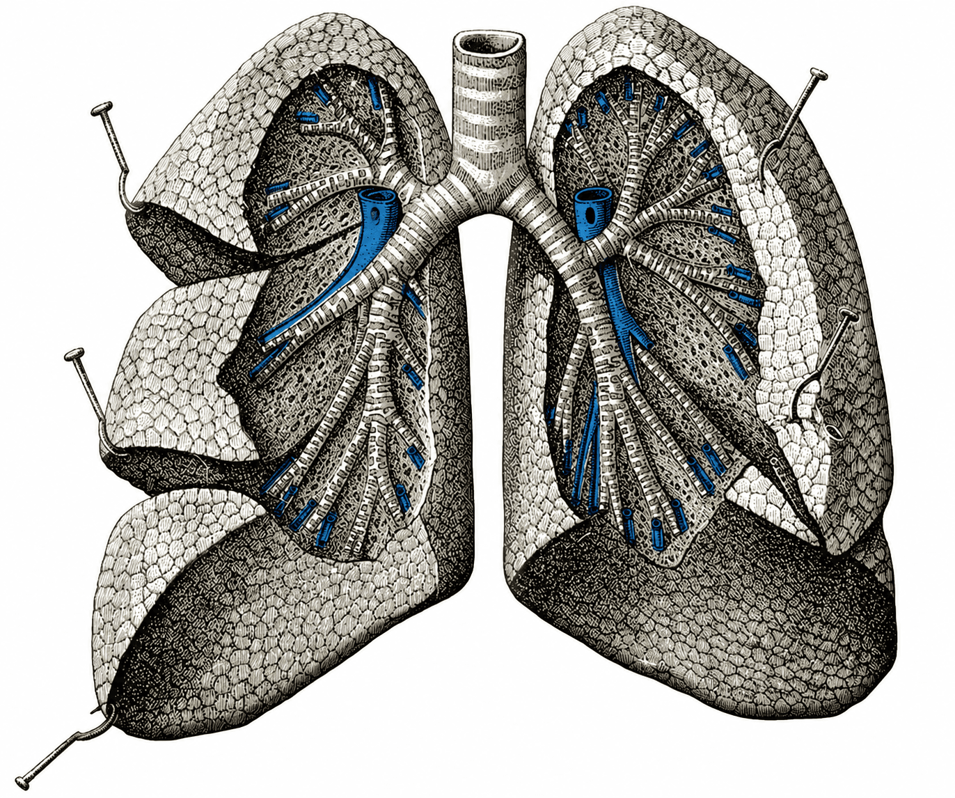
Lungs, lobes & fissures. The right lung contains upper, middle, and lower lobes. The left lung contains upper and lower lobes, with the lingula forming part of the left upper lobe.
Base anatomical illustration: public-domain image via Wikimedia Commons. Annotations added by PediAtlas.
Right Lung
- Upper lobe
- Middle lobe
- Lower lobe
The horizontal fissure separates the upper and middle lobes, while the oblique fissure separates the lower lobe from the upper and middle lobes.
Left Lung
- Upper lobe
- Lower lobe
The oblique fissure separates the upper and lower lobes.
The lingula is part of the left upper lobe and is anatomically analogous to the right middle lobe.
Bronchopulmonary Segments
Lobes can be localized even further
Each lobe is subdivided into bronchopulmonary segments supplied by segmental bronchi. You do not need to memorize every segment here. The important concept is that the bronchial tree creates progressively smaller anatomic regions that can help localize disease.
Clinical Localization
A lobe is more than an anatomy label
Descriptions such as right lower lobe pneumonia, focal atelectasis, or a localized aspiration pattern identify where in the branching respiratory system disease is occurring. Later, imaging and physical examination findings will build on this same map.
Outside the lung
Pleura & Chest Wall
The lungs do not function in isolation. Pleural membranes, the chest wall, and the diaphragm create the mechanical environment that allows the lungs to expand.
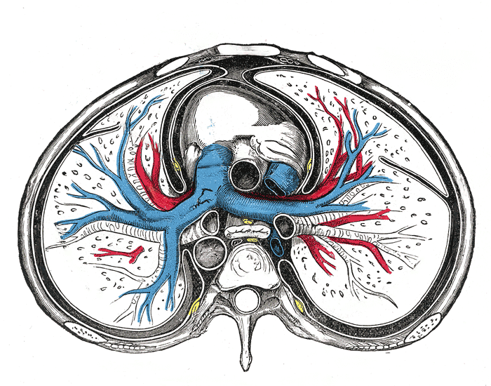
Pleura & chest wall. The visceral pleura covers the lung surface, while the parietal pleura lines the thoracic cavity. The pleural space lies between these two layers.
Hover over or select a labeled structure to highlight its anatomical location.
Base anatomical illustration: public-domain image via Wikimedia Commons. Interactive annotations added by PediAtlas.
Visceral Pleura
The visceral pleura closely covers the surface of the lung and extends into the fissures between the lobes.
Think of it as the pleural layer that travels with the lung.
Pleural Space
Between the visceral and parietal pleura is the pleural space, a potential space containing a small amount of pleural fluid.
This thin fluid layer allows the pleural surfaces to move smoothly against one another during respiration.
Parietal Pleura
The parietal pleura lines the inner surface of the thoracic cavity, including the chest wall and diaphragm.
Think of it as the pleural layer that stays with the thorax.
Respiratory Pump
The lung expands because the thorax moves
The lungs sit inside a mechanically active thorax. Movement of the diaphragm and chest wall changes thoracic volume, while the closely opposed pleural surfaces allow those movements to be transmitted to the lungs.
We will build on this relationship when we discuss intrathoracic pressure, compliance, and work of breathing in Respiratory Physiology.
Clinical Localization
Pleural disease is outside the lung itself
Localization matters. Pneumonia primarily involves the pulmonary parenchyma, while a pleural effusion places fluid in the pleural space and a pneumothorax places air there.
These processes can all impair breathing, but they occur in different anatomical compartments and therefore produce different examination, imaging, and ultrasound findings.
Clinical Pearl
Pleural pain comes from the parietal pleura
The visceral pleura does not carry pain sensation in the same way as the parietal pleura. Irritation of the parietal pleura can therefore produce the sharp, respiration-associated pain we describe clinically as pleuritic pain.
Air meets blood
Pulmonary Circulation
Follow blood from the right ventricle to the alveolar capillary bed and back to the left heart.
Section coming next.
Pediatrics changes the anatomy
Why Kids Are Different
Small airways, a compliant chest wall, developing lungs, and high metabolic demand change respiratory physiology in children.
Section coming next.
Put the map to work
Clinical Localization
Use respiratory findings to identify where in the system a problem is most likely occurring.
Section coming next.
Knowledge check
Can You Localize It?
Apply the respiratory map before moving into physiology.
Quiz coming after the teaching sections are complete.